Characterization of a neutralizing antibody that recognizes a loop region adjacent to the receptor-binding interface of the SARS-CoV-2 spike receptor-binding domain
- PMID: 38415660
- PMCID: PMC10986471
- DOI: 10.1128/spectrum.03655-23
Characterization of a neutralizing antibody that recognizes a loop region adjacent to the receptor-binding interface of the SARS-CoV-2 spike receptor-binding domain
Abstract
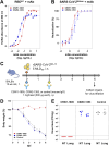
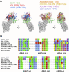
Although the global crisis caused by the coronavirus disease 2019 (COVID-19) pandemic is over, the global epidemic of the disease continues. Severe acute respiratory syndrome coronavirus-2 (SARS-CoV-2), the cause of COVID-19, initiates infection via the binding of the receptor-binding domain (RBD) of its spike protein to the human angiotensin-converting enzyme II (ACE2) receptor, and this interaction has been the primary target for the development of COVID-19 therapeutics. Here, we identified neutralizing antibodies against SARS-CoV-2 by screening mouse monoclonal antibodies and characterized an antibody, CSW1-1805, that targets a narrow region at the RBD ridge of the spike protein. CSW1-1805 neutralized several variants in vitro and completely protected mice from SARS-CoV-2 infection. Cryo-EM and biochemical analyses revealed that this antibody recognizes the loop region adjacent to the ACE2-binding interface with the RBD in both a receptor-inaccessible "down" state and a receptor-accessible "up" state and could stabilize the RBD conformation in the up-state. CSW1-1805 also showed different binding orientations and complementarity determining region properties compared to other RBD ridge-targeting antibodies with similar binding epitopes. It is important to continuously characterize neutralizing antibodies to address new variants that continue to emerge. Our characterization of this antibody that recognizes the RBD ridge of the spike protein will aid in the development of future neutralizing antibodies.IMPORTANCESARS-CoV-2 cell entry is initiated by the interaction of the viral spike protein with the host cell receptor. Therefore, mechanistic findings regarding receptor recognition by the spike protein help uncover the molecular mechanism of SARS-CoV-2 infection and guide neutralizing antibody development. Here, we characterized a SARS-CoV-2 neutralizing antibody that recognizes an epitope, a loop region adjacent to the receptor-binding interface, that may be involved in the conformational transition of the receptor-binding domain (RBD) of the spike protein from a receptor-inaccessible "down" state into a receptor-accessible "up" state, and also stabilizes the RBD in the up-state. Our mechanistic findings provide new insights into SARS-CoV-2 receptor recognition and guidance for neutralizing antibody development.
Keywords: conformational transition; monoclonal antibody; neutralizing epitope; receptor-binding domain; severe acute respiratory syndrome-coronavirus 2 (SARS-CoV-2); spike protein.
Conflict of interest statement
Yoichiro Kosaka, Yuki Miyamoto, and Tadahiro Kajita are employees of Biomatrix Inc. Koichiro Suzuki is an employee of the Research Foundation for Microbial Diseases of Osaka University. The other authors declare no competing financial interests.
Figures


Similar articles
-
Structural Basis of a Human Neutralizing Antibody Specific to the SARS-CoV-2 Spike Protein Receptor-Binding Domain.Microbiol Spectr. 2021 Oct 31;9(2):e0135221. doi: 10.1128/Spectrum.01352-21. Epub 2021 Oct 13. Microbiol Spectr. 2021. PMID: 34643438 Free PMC article.
-
Key residues of the receptor binding motif in the spike protein of SARS-CoV-2 that interact with ACE2 and neutralizing antibodies.Cell Mol Immunol. 2020 Jun;17(6):621-630. doi: 10.1038/s41423-020-0458-z. Epub 2020 May 15. Cell Mol Immunol. 2020. PMID: 32415260 Free PMC article.
-
Competitive SARS-CoV-2 Serology Reveals Most Antibodies Targeting the Spike Receptor-Binding Domain Compete for ACE2 Binding.mSphere. 2020 Sep 16;5(5):e00802-20. doi: 10.1128/mSphere.00802-20. mSphere. 2020. PMID: 32938700 Free PMC article.
-
Recognition of the SARS-CoV-2 receptor binding domain by neutralizing antibodies.Biochem Biophys Res Commun. 2021 Jan 29;538:192-203. doi: 10.1016/j.bbrc.2020.10.012. Epub 2020 Oct 10. Biochem Biophys Res Commun. 2021. PMID: 33069360 Free PMC article. Review.
-
Interactions of angiotensin-converting enzyme-2 (ACE2) and SARS-CoV-2 spike receptor-binding domain (RBD): a structural perspective.Mol Biol Rep. 2023 Mar;50(3):2713-2721. doi: 10.1007/s11033-022-08193-4. Epub 2022 Dec 23. Mol Biol Rep. 2023. PMID: 36562937 Free PMC article. Review.
Cited by
-
Humoral Immune Response to SARS-CoV-2 Spike Protein Receptor-Binding Motif Linear Epitopes.Vaccines (Basel). 2024 Mar 22;12(4):342. doi: 10.3390/vaccines12040342. Vaccines (Basel). 2024. PMID: 38675725 Free PMC article.
-
SARS-CoV-2 BA.2.86 is susceptible to the neutralizing antibody MO11 targeting subdomain 1 despite the E554K mutation near the epitope.J Virol. 2025 Feb 25;99(2):e0138924. doi: 10.1128/jvi.01389-24. Epub 2025 Jan 22. J Virol. 2025. PMID: 39840983 Free PMC article. No abstract available.
References
-
- Zhu N, Zhang D, Wang W, Li X, Yang B, Song J, Zhao X, Huang B, Shi W, Lu R, Niu P, Zhan F, Ma X, Wang D, Xu W, Wu G, Gao GF, Tan W, China Novel Coronavirus Investigating and Research Team . 2020. A novel coronavirus from patients with pneumonia in China, 2019. N Engl J Med 382:727–733. doi:10.1056/NEJMoa2001017 - DOI - PMC - PubMed
-
- WHO . WHO coronavirus (COVID-19) dashboard with vaccination data. Available from: https://covid19.who.int
MeSH terms
Substances
Grants and funding
- JP16H06429, JP16K21723, JP16H06432/Ministry of Education, Culture, Sports, Science and Technology (MEXT)
- JP16H06429, JP16K21723, JP16H06434/Ministry of Education, Culture, Sports, Science and Technology (MEXT)
- JP22H02521/MEXT | Japan Society for the Promotion of Science (JSPS)
- JP21K15042/MEXT | Japan Society for the Promotion of Science (JSPS)
- JP21H02736/MEXT | Japan Society for the Promotion of Science (JSPS)
- JP25K000013/MEXT | Japan Society for the Promotion of Science (JSPS)
- JP20K22630/MEXT | Japan Society for the Promotion of Science (JSPS)
- JP223fa627002, JP22am0401030, JP23fk0108659, JP20jk0210021, JP22gm1610010, JP19fk0108113/Japan Agency for Medical Research and Development (AMED)
- JP223fa627002/Japan Agency for Medical Research and Development (AMED)
- JP19fk0108113, JP20fk0108281, JP20pc0101047/Japan Agency for Medical Research and Development (AMED)
- JP20fk0108401, JP21fk0108493/Japan Agency for Medical Research and Development (AMED)
- JP21am0101117, JP17pc0101020/Japan Agency for Medical Research and Development (AMED)
- JPMJOP1861/MEXT | Japan Science and Technology Agency (JST)
- JPMJMS2025/MEXT | Japan Science and Technology Agency (JST)
LinkOut - more resources
Full Text Sources
Medical
Molecular Biology Databases
Miscellaneous

