

Effect of Potassium Permanganate on Staphylococcal Isolates Derived from the Skin of Patients with Atopic Dermatitis
- PMID: 38415865
- PMCID: PMC10916795
- DOI: 10.2340/actadv.v104.18642
Effect of Potassium Permanganate on Staphylococcal Isolates Derived from the Skin of Patients with Atopic Dermatitis
Abstract
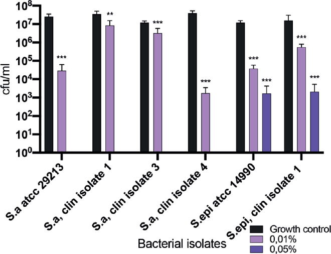
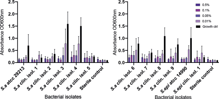
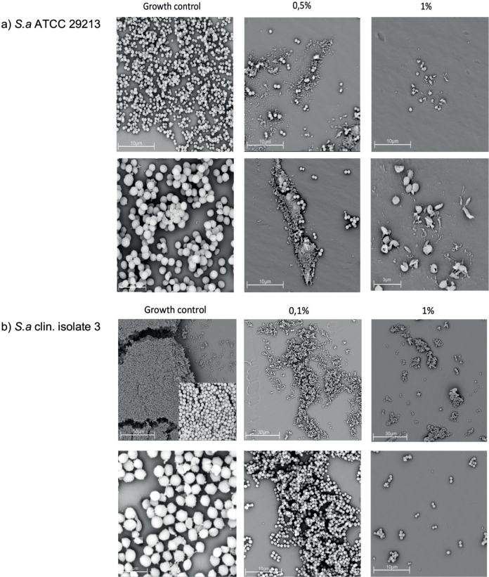
In atopic dermatitis (AD), Staphylococcus aureus frequently colonizes lesions, leading to superinfections that can then lead to exacerbations. The presence of biofilm-producing isolates has been associated with worsening of the disease. Potassium permanganate is used as a topical treatment of infected eczema, blistering conditions, and wounds. Little is known of its effects against microbes in AD skin. The aim of this study was to explore antibacterial and antibiofilm properties of potassium permanganate against staphylococcal isolates derived from AD skin. Viable count and radial diffusion assays were used to investigate antibacterial effects of potassium permanganate against planktonic staphylococcal isolates. The antibiofilm effects were assessed using biofilm assays and scanning electron microscopy. The Staphylococcus aureus isolates were completely killed when exposed to 0.05% of potassium permanganate. In concentrations of 0.01%, potassium permanganate inhibited bacterial biofilm formation. Eradication of established staphylococcal biofilm was observed in concentrations of 1%. Electron microscopy revealed dense formations of coccoidal structures in growth control and looser formations of deformed bacteria when exposed to potassium permanganate. This suggests antibacterial and antibiofilm effects of potassium permanganate against staphylococcal isolates derived from AD skin, when tested in vitro, and a potential role in the treatment of superinfected AD skin.
Conflict of interest statement
AS has received speaker honoraria and consulting fees from AbbVie, LEO Pharma, Pfizer, and Sanofi. Payments were made to AS’s institution. AS has been an investigator for AbbVie.
Figures

Similar articles
-
Antibacterial and antibiofilm effects of sodium hypochlorite against Staphylococcus aureus isolates derived from patients with atopic dermatitis.Br J Dermatol. 2017 Aug;177(2):513-521. doi: 10.1111/bjd.15410. Epub 2017 Jul 3. Br J Dermatol. 2017. PMID: 28238217
-
Biofilm propensity of Staphylococcus aureus skin isolates is associated with increased atopic dermatitis severity and barrier dysfunction in the MPAACH pediatric cohort.Allergy. 2021 Jan;76(1):302-313. doi: 10.1111/all.14489. Epub 2020 Aug 9. Allergy. 2021. PMID: 32640045 Free PMC article.
-
Skin colonization by Staphylococcus aureus in patients with eczema and atopic dermatitis and relevant combined topical therapy: a double-blind multicentre randomized controlled trial.Br J Dermatol. 2006 Oct;155(4):680-7. doi: 10.1111/j.1365-2133.2006.07410.x. Br J Dermatol. 2006. PMID: 16965415 Clinical Trial.
-
Skin microbiome dysbiosis and the role of Staphylococcus aureus in atopic dermatitis in adults and children: A narrative review.J Eur Acad Dermatol Venereol. 2023 Jun;37 Suppl 5:3-17. doi: 10.1111/jdv.19125. J Eur Acad Dermatol Venereol. 2023. PMID: 37232427 Review.
-
Staphylococcus aureus colonization in atopic dermatitis and its therapeutic implications.Br J Dermatol. 1998 Dec;139 Suppl 53:13-6. doi: 10.1046/j.1365-2133.1998.1390s3013.x. Br J Dermatol. 1998. PMID: 9990408 Review.
References
-
- Weidinger, S. and N. Novak. Atopic dermatitis. Lancet 2016; 387: 1109–1122. - PubMed
-
- Silverberg JI, Hanifin JM. Adult eczema prevalence and associations with asthma and other health and demographic factors: a US population-based study. J Allergy Clin Immunol 2013; 132: 1132–1138. - PubMed
-
- Allen HB, Vaze ND, Choi C, Hailu T, Tulbert BH, Cusack C, et al. . The presence and impact of biofilm-producing staphylococci in atopic dermatitis. JAMA Dermatol 2014; 150: 260–265. - PubMed
-
- Breuer K, S HA, Kapp A, Werfel T. Staphylococcus aureus: colonizing features and influence of an antibacterial treatment in adults with atopic dermatitis. Br J Dermatol 2002; 147: 55–61. - PubMed
MeSH terms
Substances
LinkOut - more resources
Full Text Sources
Medical
Molecular Biology Databases
