. 2024 Jun 1;17(6):dmm050358.
doi: 10.1242/dmm.050358. Epub 2024 Feb 28.

Smad4 restricts injury-provoked biliary proliferation and carcinogenesis

Affiliations

Smad4 restricts injury-provoked biliary proliferation and carcinogenesis

William B Alexander et al. Dis Model Mech. .

Abstract

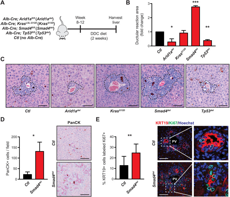
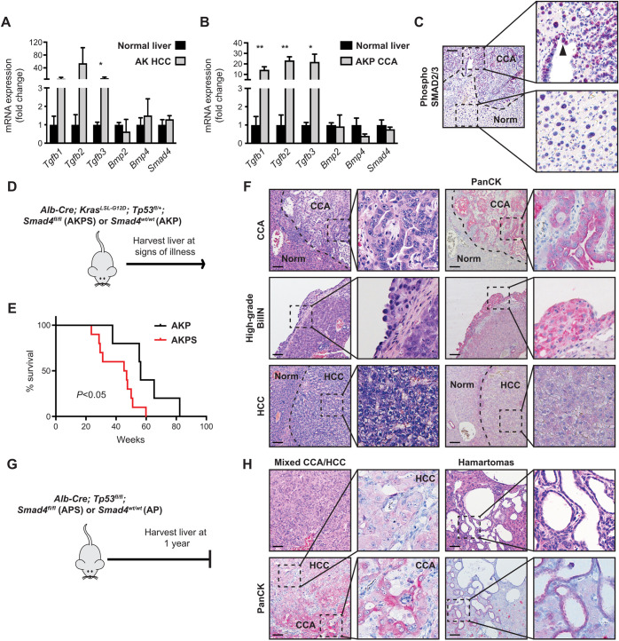
Cholangiocarcinoma (CCA) is a deadly and heterogeneous type of cancer characterized by a spectrum of epidemiologic associations as well as genetic and epigenetic alterations. We seek to understand how these features inter-relate in the earliest phase of cancer development and through the course of disease progression. For this, we studied murine models of liver injury integrating the most commonly occurring gene mutations of CCA - including Kras, Tp53, Arid1a and Smad4 - as well as murine hepatobiliary cancer models and derived primary cell lines based on these mutations. Among commonly mutated genes in CCA, we found that Smad4 functions uniquely to restrict reactive cholangiocyte expansion to liver injury through restraint of the proliferative response. Inactivation of Smad4 accelerates carcinogenesis, provoking pre-neoplastic biliary lesions and CCA development in an injury setting. Expression analyses of Smad4-perturbed reactive cholangiocytes and CCA lines demonstrated shared enriched pathways, including cell-cycle regulation, MYC signaling and oxidative phosphorylation, suggesting that Smad4 may act via these mechanisms to regulate cholangiocyte proliferation and progression to CCA. Overall, we showed that TGFβ/SMAD4 signaling serves as a critical barrier restraining cholangiocyte expansion and malignant transformation in states of biliary injury.

Keywords: Biliary epithelium; Cholangiocarcinoma; Methylation; Murine models of liver injury; TGFβ/SMAD4.


Conflict of interest statement

Competing interests The authors declare no competing or financial interests.

Figures

Fig. 1.
Fig. 1.
Smad4 suppresses cholangiocyte proliferation in response to injury. (A) Generation of compound mutant mice carrying Alb-Cre and Arid1afl/fl (Arid1adel), KrasLSL-G12D (KrasG12D), Smad4fl/fl (Smad4del) or Tp53fl/fl (Tp53del). Experimental and control cohorts [no Alb-Cre, (Ctl)] were put on a DDC diet for 2 weeks and their livers were harvested, n=3-4 mice per cohort. (B) Quantification of the ductular reaction area of the experimental cohorts compared to the control cohort as outlined in A, showing Smad4del with a significant expansion of the biliary compartment. (C) Representative images of ductular reactions (surrounded by dashed lines), magnified 200×. (D) Quantification of reactive cholangiocytes (identified as PanCK+) in Smad4del and Ctl livers demonstrating more reactive cholangiocytes in the DDC-injured Smad4del livers compared to that of Ctl. Three random images per mouse (magnified 100×) were used for quantification. Representative ductular reactions are shown on the right. (E) Quantification of proliferating cholangiocytes (KRT19+ and Ki67+) in Smad4del and Ctl livers revealing more proliferative cholangiocytes in DDC-injured Smad4del livers compared to that of Ctl. For quantification, three random portal fields per mouse were aggregated by genotype. Representative images (magnified 100×) are shown; boxed areas are shown enlarged on the right. *P<0.05, **P<0.01, ***P<0.0001; scale bars: 50 µm (C) and 100 µm (D,E).
Fig. 2.
Fig. 2.
Smad4 is involved in the regulation of pathways associated with proliferation, metabolism and inflammation. (A) Schematic outlining the strategy to isolate and sequence reactive cholangiocytes. Livers are enzymatically digested and flow-sorted to isolate the EPCAM-positive/CD45-negative/Ter119-negative (EPCAM+/CD45-/Ter119-) population from which RNA had been prepared for RNA sequencing. Representative FACS plots are shown on the left. Boxed areas enclose cells of interest (P5 = CD45/Ter119 cells, EpCAM = EpCAM+ cells). n=5 per cohort. (B) Heat map showing the top 50 most significantly upregulated and downregulated genes in Smad4del-perturbed reactive cholangiocytes. (C) Gene set enrichment analysis (GSEA) of genes differentially expressed in Smad4del-perturbed reactive cholangiocytes. Enrichment plots from the Hallmarks Gene Set Collection are shown for the top five gene sets most positively correlated (top row) in genes upregulated in Smad4del cells or most negatively correlated (bottom row) in genes downregulated in Smad4del cells. Ox-Phos, oxidative phosphorylation; EMT, epithelial mesenchymal transition; NES, normalized enrichment score.
Fig. 3.
Fig. 3.
Smad4 suppresses cancer progression in the AKP and AP hepatobiliary cancer models. (A) mRNA expression levels assessed by qPCR of TGFβ family-related genes in (A) AK mouse model-derived HCC (AK HCC) or (B) AKP mouse model-derived CCA (AKP CCA) tissue compared to normal liver tissue showing increased expression of TGFβ ligands, indicating activation of the TGFβ pathway. Expression was normalized to Rhoa, n=3 mice per cohort. (C) Immunohistochemistry image of phosphorylated (phospho)-SMAD2 in AKP CCA tissue and adjacent normal (Norm) tissue (magnified 100×). Boxed areas are shown enlarged on the right, with CCA tissue at the top and normal tissue at the bottom. Arrowhead shows positive phospho-SMAD2 nuclei in malignant epithelium. (D) Compound mutant mice carrying Alb-Cre; KrasLSL-G12D; Tp53fl/+ and either Smad4fl/fl or Smad4wt/wt alleles (AKPS or AKP mouse models, respectively) were followed for survival (n=5–10 mice per cohort). (E) Kaplan–Meier plot comparing survival curves of AKPS (red) and AKP (black) mice, showing decreased overall survival of the AKPS compared to the AKP cohort (P<0.05). (F) Representative histology and immunohistochemistry images of AKPS CCA, high-grade BilIN and HCC (magnified 100×). Boxed areas are shown enlarged to the right. Staining of reactive cholangiocytes (PanCK) highlights malignant biliary epithelium. (G) Compound mutant mice carrying Alb-Cre;Tp53fl/fl and Smad4fl/fl or Smad4wt/wt alleles (APS or AP mouse models, respectively) were followed and sacrificed at one year of age, n=8–15 mice per cohort. (H) Representative histology images of APS mixed CCA/HCC and hamartomas (magnified 100×). Boxed areas are shown enlarged to the right. Staining of PanCK highlights biliary differentiation. *P<0.05, **P<0.01, Scale bars: 50 µm.
Fig. 4.
Fig. 4.
Pathway analyses, suggesting conserved functions of Smad4 in CCA and reactive cholangiocytes. (A) Plotted are the growth curves of two CCA cell lines (#1 and #2) derived from liver tumors from mice in the AKPS cohort expressing only Smad4 (red) or empty vector (EV; black), and Smad4 or EV plus treatment with TGFβ (gray or orange, respectively), showing growth arrest/inhibition with TGFβ treatment following reintroduction of Smad4 to both AKPS #1 and #2. (B) Western blot confirming restoration of Smad4 (S4) protein levels in the two AKPS lines assessed in A. (C) Heat map showing the top 50 most significantly up- and downregulated genes in Smad4-restored CCA cell lines. (D) Individual and shared gene sets enriched with loss of Smad4 signaling (top) and with intact Smad4 signaling (bottom) in AKPS CCA cell lines (right) compared to those enriched in reactive cholangiocytes (left).
Fig. 5.
Fig. 5.
Smad4 is a determinant of DNA methylation. (A) Histograms indicating the frequency of differentially methylated regions binned according to changes in DNA methylation (DNAme; x-axis) in the two AKPS mouse model-derived CCA lines described in Fig.4. Increases (red) and decreases (blue) of DNAme were scored for AKPS #2 versus AKPS #1 (top) and for Smad4 re-expression empty vector (EV) (bottom). (B) Boxplots indicate the distance (in kb) of differentially methylated regions relative to the nearest transcriptional start site (TSS). Regions from ‘Decreased DNAme Smad4 vs. EV’ were found at a greater distance from the nearest TSS, suggesting a differential effect of Smad4 on DNAme distal to promoter regions. (C) Differentially methylated regions were analyzed based on sample type (AKPS #2 vs AKPS #1; two pie charts on left) or Smad4 re-expression [Smad4 vs. EV; two pie charts on right) and further determined based on increased or decreased DNAme. Regions were then annotated and color-coded based on genetic context.

Similar articles

Cited by

References

    1. Banales, J. M., Cardinale, V., Carpino, G., Marzioni, M., Andersen, J. B., Invernizzi, P., Lind, G. E., Folseraas, T., Forbes, S. J., Fouassier, L.et al. (2016). Expert consensus document: Cholangiocarcinoma: current knowledge and future perspectives consensus statement from the European Network for the Study of Cholangiocarcinoma (ENS-CCA). Nat. Rev. Gastroenterol. Hepatol. 13, 261-280. 10.1038/nrgastro.2016.51 - DOI - PubMed
    1. Bardeesy, N., Cheng, K. H., Berger, J. H., Chu, G. C., Pahler, J., Olson, P., Hezel, A. F., Horner, J., Lauwers, G. Y., Hanahan, D.et al. (2006). Smad4 is dispensable for normal pancreas development yet critical in progression and tumor biology of pancreas cancer. Genes Dev. 20, 3130-3146. 10.1101/gad.1478706 - DOI - PMC - PubMed
    1. Bolger, A. M., Lohse, M. and Usadel, B. (2014). Trimmomatic: a flexible trimmer for Illumina sequence data. Bioinformatics 30, 2114-2120. 10.1093/bioinformatics/btu170 - DOI - PMC - PubMed
    1. Cardenas, H., Vieth, E., Lee, J., Segar, M., Liu, Y., Nephew, K. P. and Matei, D. (2014). TGF-beta induces global changes in DNA methylation during the epithelial-to-mesenchymal transition in ovarian cancer cells. Epigenetics 9, 1461-1472. 10.4161/15592294.2014.971608 - DOI - PMC - PubMed
    1. Chan-On, W., Nairismagi, M. L., Ong, C. K., Lim, W. K., Dima, S., Pairojkul, C., Lim, K. H., Mcpherson, J. R., Cutcutache, I., Heng, H. L.et al. (2013). Exome sequencing identifies distinct mutational patterns in liver fluke-related and non-infection-related bile duct cancers. Nat. Genet. 45, 1474-1478. 10.1038/ng.2806 - DOI - PubMed

Substances