

High-Resolution Profiling of Human Vocal Fold Cellular Landscapes With Single-Nuclei RNA Sequencing
- PMID: 38415934
- PMCID: PMC12279057
- DOI: 10.1002/lary.31334
High-Resolution Profiling of Human Vocal Fold Cellular Landscapes With Single-Nuclei RNA Sequencing
Abstract
Introduction: The function of the vocal folds (VFs) is determined by the phenotype, abundance, and distribution of differentiated cells within specific microenvironments. Identifying this histologic framework is crucial in understanding laryngeal disease. A paucity of studies investigating VF cellular heterogeneity has been undertaken. Here, we examined the cellular landscape of human VFs by utilizing single-nuclei RNA-sequencing.
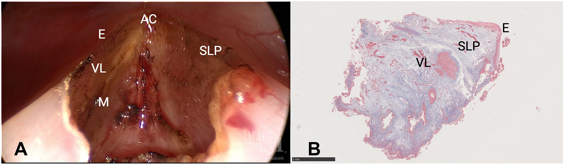
Methods: Normal true VF tissue was excised from five patients undergoing pitch elevation surgery. Tissue was snap frozen in liquid nitrogen and subjected to cellular digestion and nuclear extraction. Nuclei were processed for single-nucleus sequencing using the 10X Genomics Chromium platform. Sequencing reads were assembled using cellranger and analyzed with the scanpy package in python.
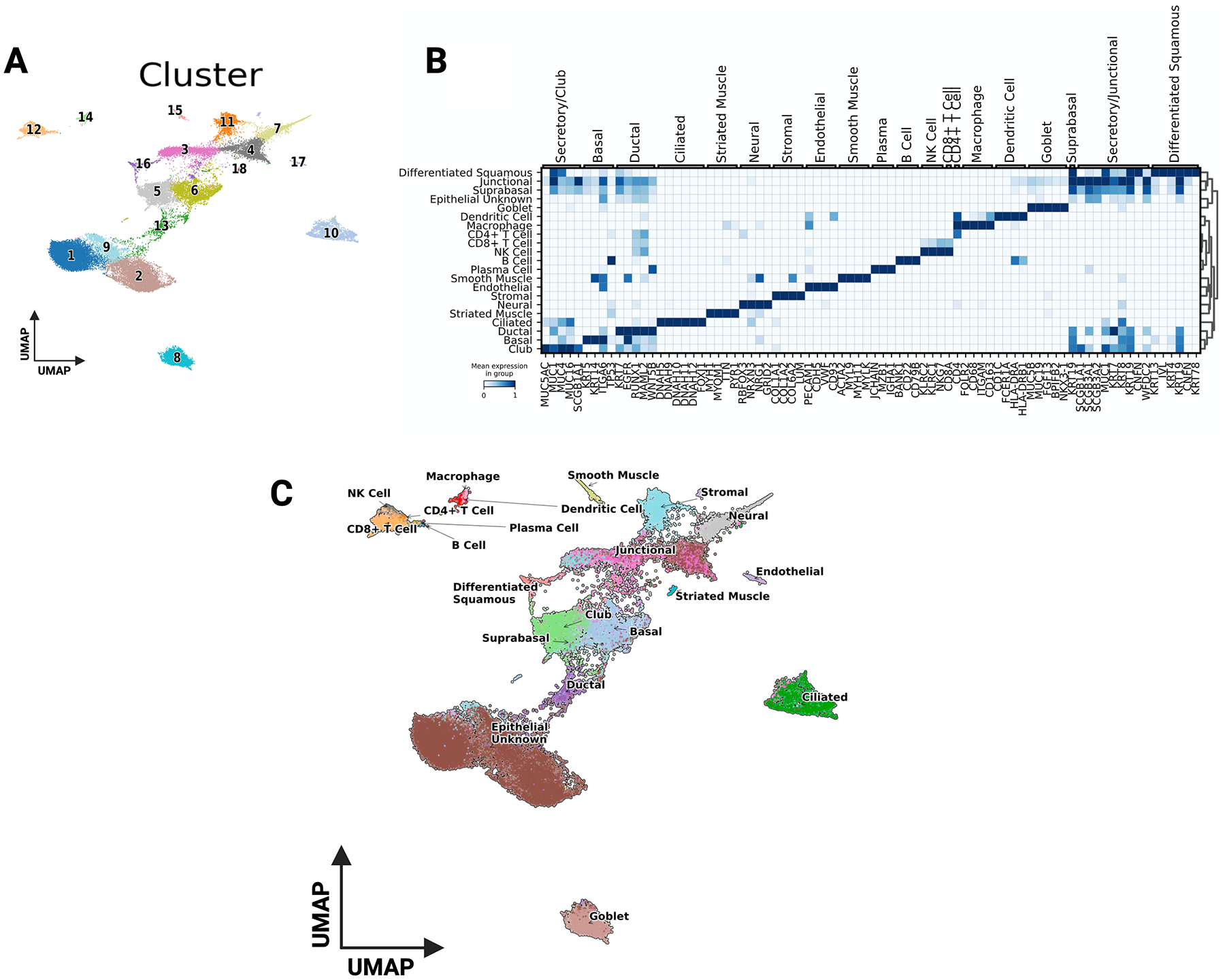
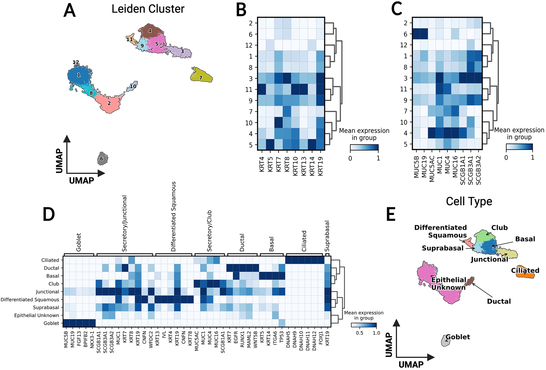
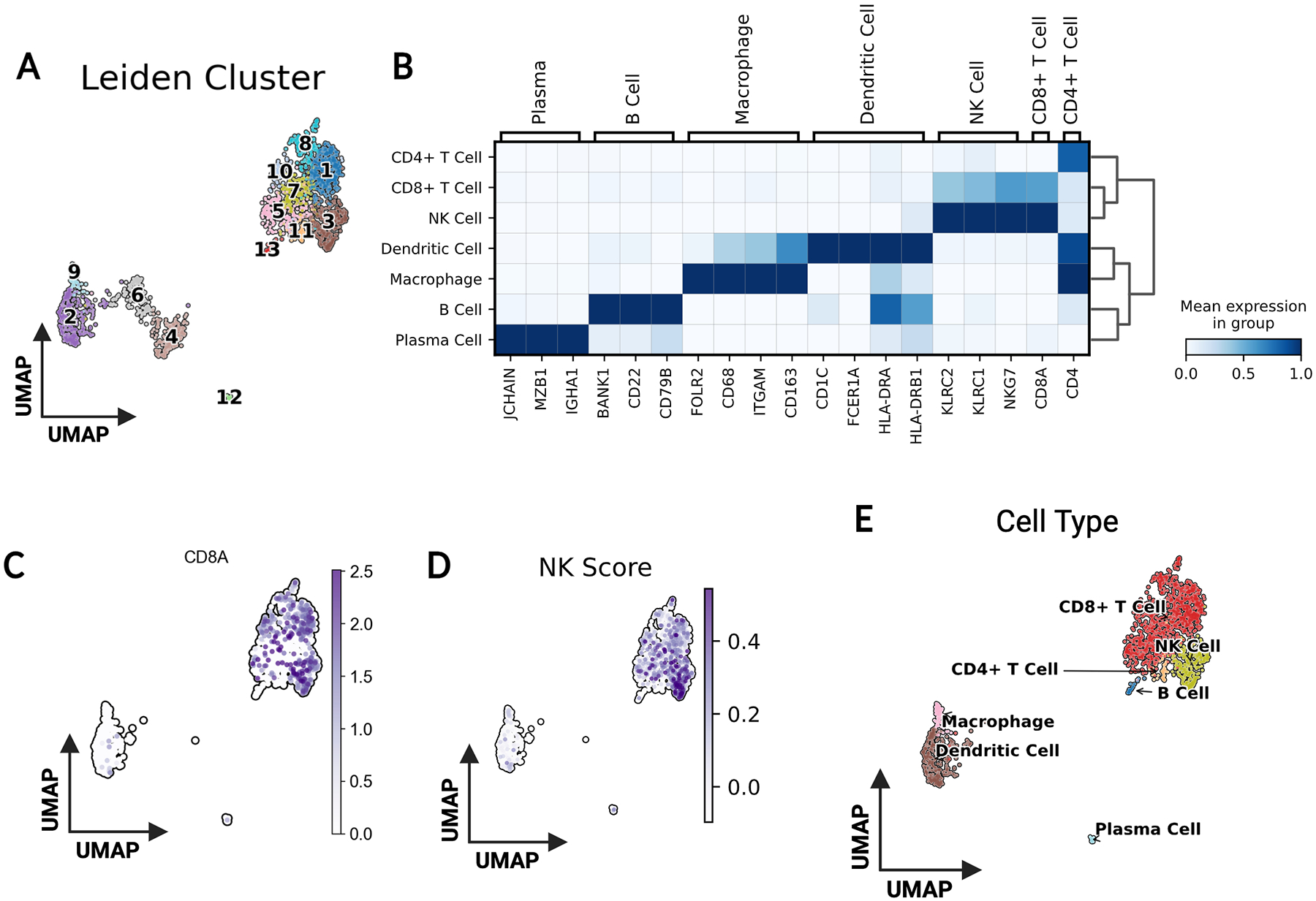
Results: RNA sequencing revealed 18 global cell clusters. While many were of epithelial origin, expected cell types, such as fibroblasts, immune cells, muscle cells, and endothelial cells were present. Subcluster analysis defined unique epithelial, immune, and fibroblast subpopulations.
Conclusion: This study evaluated the cellular heterogeneity of normal human VFs by utilizing single-nuclei RNA-sequencing. With further confirmation through additional spatial sequencing and microscopic imaging, a novel cellular map of the VFs may provide insight into new cellular targets for VF disease.
Level of evidence: NA Laryngoscope, 134:3193-3200, 2024.
Keywords: cellular architecture; single nuclei RNA sequencing; vocal fold.
© 2024 The American Laryngological, Rhinological and Otological Society, Inc.
Conflict of interest statement
Figures
Similar articles
-
Single-cell analysis comparing early-stage oocytes from fresh and slow-frozen/thawed human ovarian cortex reveals minimal impact of cryopreservation on the oocyte transcriptome.Hum Reprod. 2025 Apr 1;40(4):683-694. doi: 10.1093/humrep/deaf009. Hum Reprod. 2025. PMID: 39919251
-
Can a Liquid Biopsy Detect Circulating Tumor DNA With Low-passage Whole-genome Sequencing in Patients With a Sarcoma? A Pilot Evaluation.Clin Orthop Relat Res. 2025 Jan 1;483(1):39-48. doi: 10.1097/CORR.0000000000003161. Epub 2024 Jun 21. Clin Orthop Relat Res. 2025. PMID: 38905450
-
New insights for precision treatment of glioblastoma from analysis of single-cell lncRNA expression.J Cancer Res Clin Oncol. 2021 Jul;147(7):1881-1895. doi: 10.1007/s00432-021-03584-9. Epub 2021 Mar 11. J Cancer Res Clin Oncol. 2021. PMID: 33693962 Free PMC article.
-
Signs and symptoms to determine if a patient presenting in primary care or hospital outpatient settings has COVID-19.Cochrane Database Syst Rev. 2022 May 20;5(5):CD013665. doi: 10.1002/14651858.CD013665.pub3. Cochrane Database Syst Rev. 2022. PMID: 35593186 Free PMC article.
-
The effect of sample site and collection procedure on identification of SARS-CoV-2 infection.Cochrane Database Syst Rev. 2024 Dec 16;12(12):CD014780. doi: 10.1002/14651858.CD014780. Cochrane Database Syst Rev. 2024. PMID: 39679851 Free PMC article.
Cited by
-
Single-cell atlas of healthy vocal folds and cellular function in the endothelial-to-mesenchymal transition.Cell Prolif. 2024 Dec;57(12):e13723. doi: 10.1111/cpr.13723. Epub 2024 Sep 8. Cell Prolif. 2024. PMID: 39245637 Free PMC article.
References
-
- Chhetri DK, Head C, Revazova E, Hart S, Bhuta S, Berke GS. Lamina propria replacement therapy with cultured autologous fibroblasts for vocal fold scars. Otolaryngol Head Neck Surg 2004; 131:864–870. - PubMed
MeSH terms
Grants and funding
LinkOut - more resources
Full Text Sources
Research Materials
