. 2024 Apr 15;212(8):1307-1318.
doi: 10.4049/jimmunol.2300426.

Plitidepsin as an Immunomodulator against Respiratory Viral Infections

Affiliations

Plitidepsin as an Immunomodulator against Respiratory Viral Infections

Alejandro Losada et al. J Immunol. .

Abstract

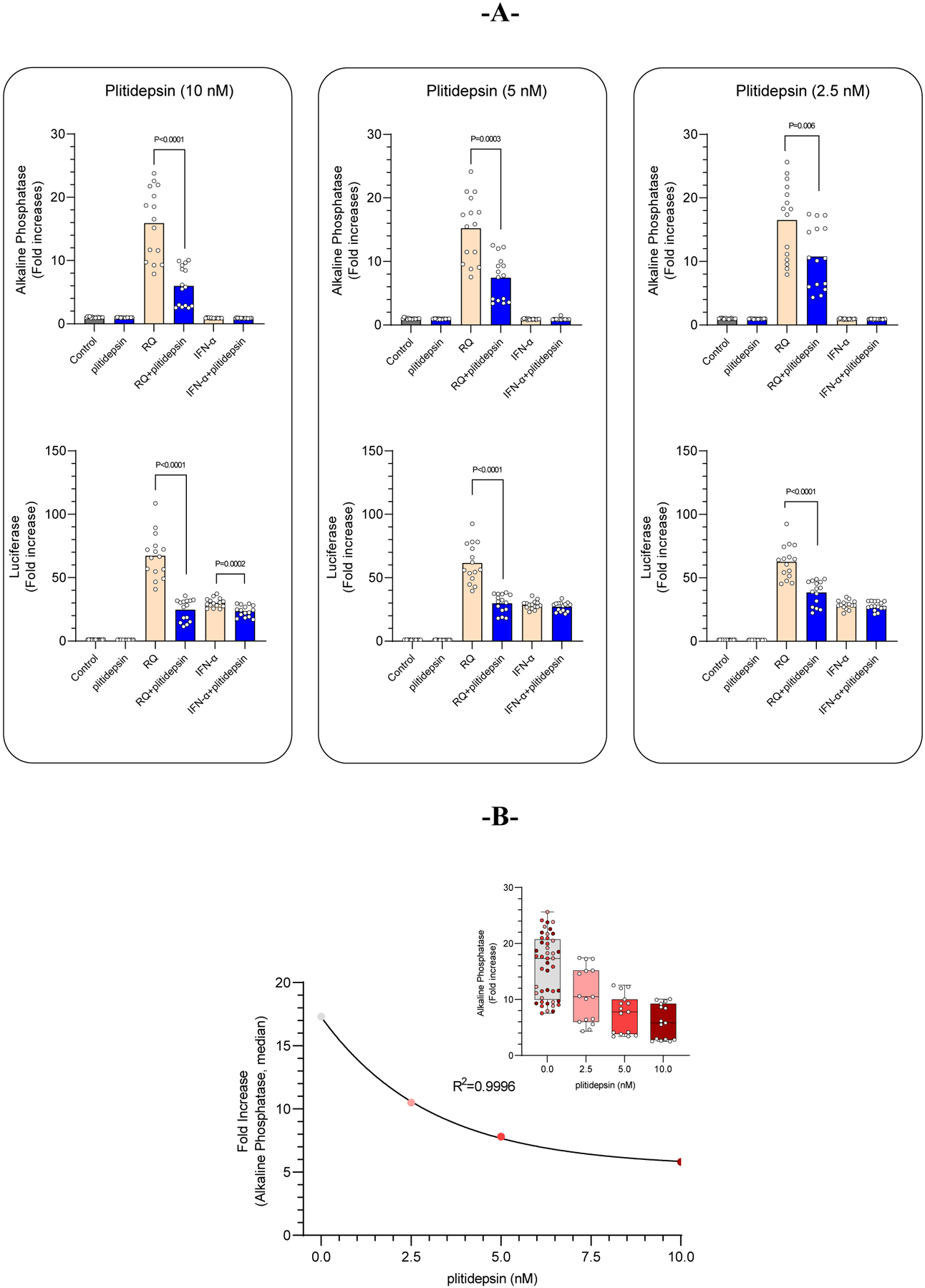
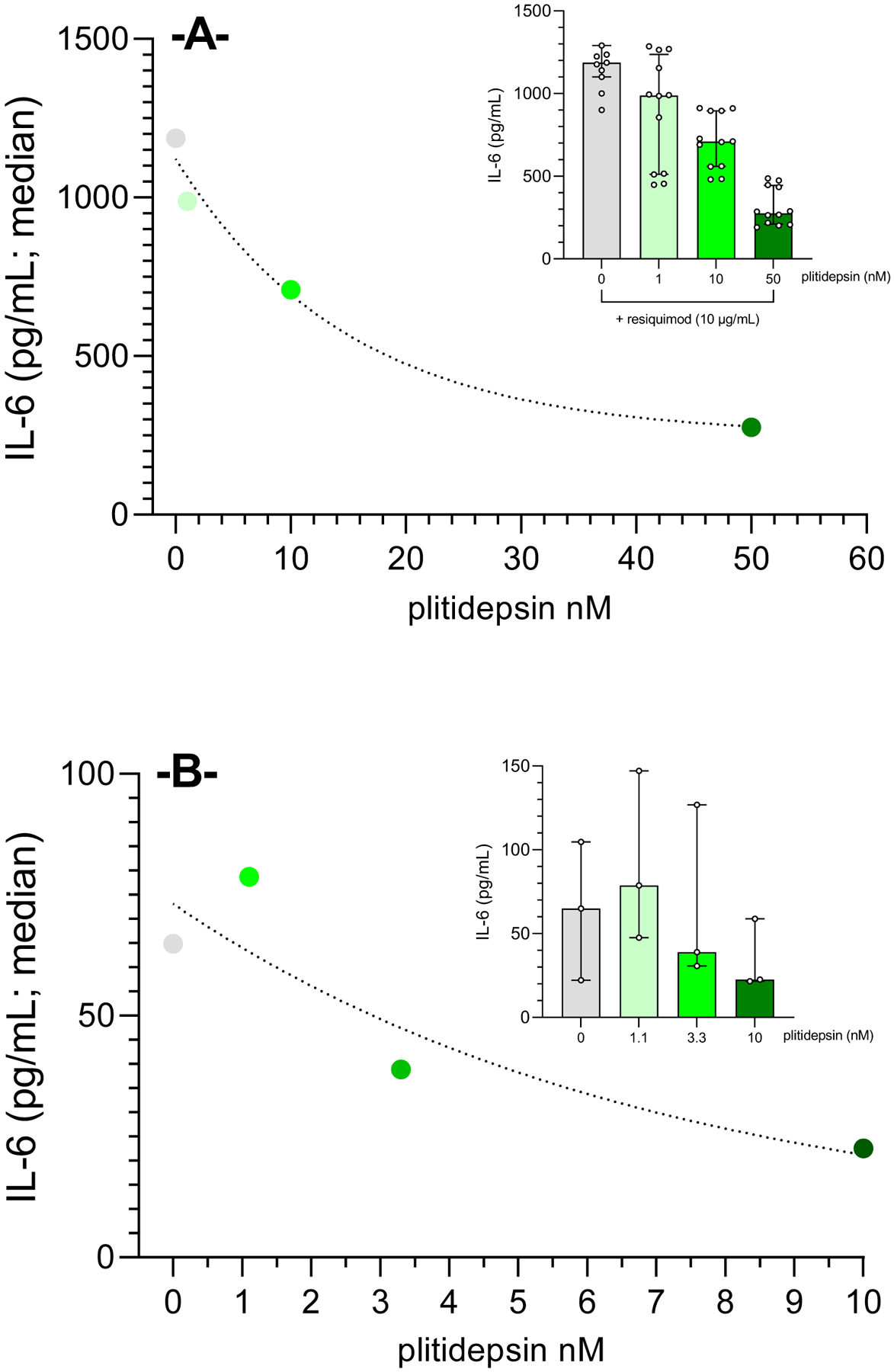
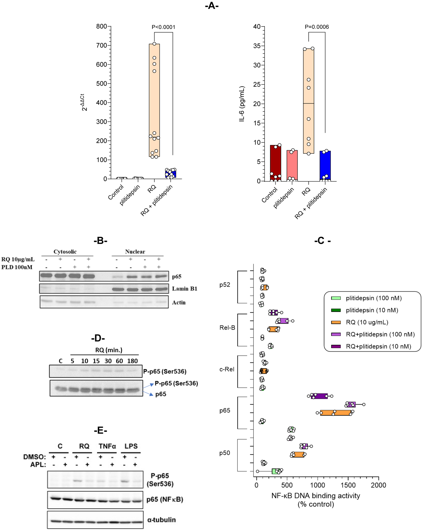
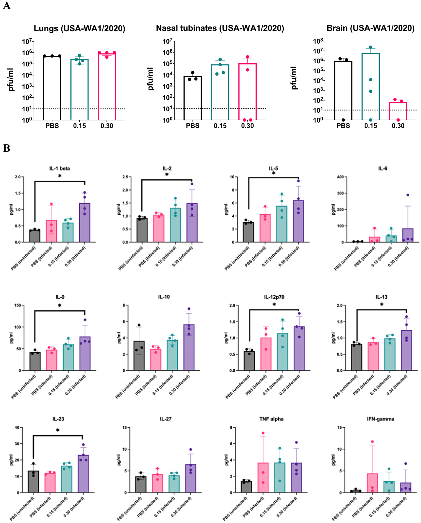
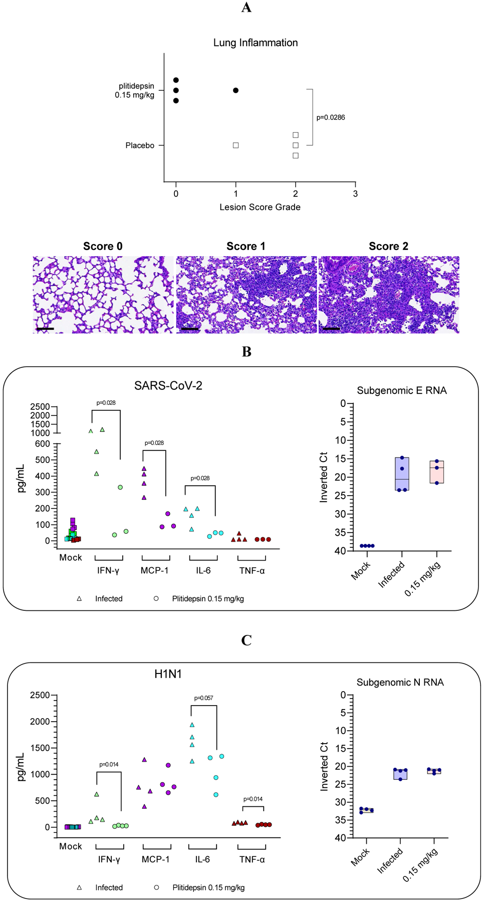
Plitidepsin is a host-targeted compound known for inducing a strong anti-SARS-CoV-2 activity, as well as for having the capacity of reducing lung inflammation. Because IL-6 is one of the main cytokines involved in acute respiratory distress syndrome, the effect of plitidepsin in IL-6 secretion in different in vitro and in vivo experimental models was studied. A strong plitidepsin-mediated reduction of IL-6 was found in human monocyte-derived macrophages exposed to nonproductive SARS-CoV-2. In resiquimod (a ligand of TLR7/8)-stimulated THP1 human monocytes, plitidepsin-mediated reductions of IL-6 mRNA and IL-6 levels were also noticed. Additionally, although resiquimod-induced binding to DNA of NF-κB family members was unaffected by plitidepsin, a decrease in the regulated transcription by NF-κB (a key transcription factor involved in the inflammatory cascade) was observed. Furthermore, the phosphorylation of p65 that is required for full transcriptional NF-κB activity was significantly reduced by plitidepsin. Moreover, decreases of IL-6 levels and other proinflammatory cytokines were also seen in either SARS-CoV-2 or H1N1 influenza virus-infected mice, which were treated at low enough plitidepsin doses to not induce antiviral effects. In summary, plitidepsin is a promising therapeutic agent for the treatment of viral infections, not only because of its host-targeted antiviral effect, but also for its immunomodulatory effect, both of which were evidenced in vitro and in vivo by the decrease of proinflammatory cytokines.


Figures

Figure 1.
Figure 1.. Plitidepsin modulates the NF-κB-regulated transcription.
Resiquimod (10 μg/mL; NF-κB signaling) or Interferon-α (20 ng/mL; Interferon signaling) -challenged THP1-Dual cells were incubated with increasing concentrations of plitidepsin for 18 hours. (A) Only a marginal plitidepsin-mediated effect was observed in luciferase levels (a marker of the interferon-regulated transcription) in the interferon-challenged THP1- Dual cell supernatants. However, a statistically significant reduction of alkaline phosphatase (a marker of NF-κB-regulated transcription) levels were quantified in the supernatants of resiquimod-challenged THP1-Dual cells, with plitidepsin-mediated inhibition of NF-κB-regulated transcription being specific and concentration-dependent (B).
Figure 2.
Figure 2.. Plitidepsin-mediated reduction of IL-6 secretion in THP-1 human monocytes and primary macrophages.
IL-6 levels quantified in the supernatants of resiquimod-challenged THP1-Dualmonocytes (A) and human monocyte-derived macrophages (MDM) exposed to non-productive SARS-CoV-2 (B) show a very strong concentration-dependent reduction when exposed to increasing concentrations of plitidepsin (THP-1 cells: 1, 10 and 50 nM for 16 h; MDM: 1.1, 3.3 and 10 nM for 24 h). Results show median values of n=12 and n=3 for THP-1 and MDM, respectively. Inlets display scatter dot plot for individual samples obtained in each experiment.
Figure 3.
Figure 3.. Plitidepsin regulates the NF-κB signaling pathway.
(A) THP-1 cells were co-treated with 100 nM of plitidepsin and 10 μg/mL RQ for 6 hours or only with plitidepsin or RQ. IL-6 RNA expression (left panel) was analyzed by qPCR and secreted IL-6 (right panel) were analyzed by ELISA. Data presented are the median of three independent experiments performed in quadruplicate. (B) THP-1 cells were stimulated with RQ (10 μg/mL) alone or in combination with plitidepsin (100 nM) for 1 hour, and nuclear and cytoplasmic fractions were obtained and analyzed by immunoblotting. Western blots revealed p65 nuclear import. The purity of the fractions was controlled with an Ab recognizing the nuclear Lamin B1 protein. (C) Binding of NF-kB family members (p50, p65, c-Rel, RelB and p52) to a specific double stranded DNA sequence containing the NF-kβ consensus binding site was quantified. THP1 cells were treated with RQ (10 μg/mL) alone or in combination with plitidepsin (10nM and 100 nM) for 2 hours. Nuclear extracts were obtained and tested for NF-κB binding. The data are the median of three independent experiments performed in duplicate. (D) THP-1 cells were stimulated for the indicated periods with RQ. Phosphorylation of p65 (Ser536) and total p65 were analyzed by Western blot. (E) Immunoblot assay showing p65 and phosphorylation of p65 (Ser536) in THP1 cells after stimulation with RQ (10 μg/mL), TNF-α (100 ng/mL) and LPS (10 μg/mL) with or without plitidepsin (100 nM) for 1 hour. Tubulin was analyzed as loading control.
Figure 4.
Figure 4.. Anti-inflammatory cytokine milieu in the lung induced by plitidepsin.
(A) Virus titers determined by plaque assay at 5 days post infection in lung, nasal turbinates and brain of hACE2 transgenic mice infected with 104 PFU of USA-WA1/2020 SARS-CoV-2 and treated with 0.15 or 0.30 mg/kg of plitidepsin for three consecutive days or placebo. Every dot represents an individual mouse. (B) Cytokine profile in cleared lung homogenates from SARS-CoV-2 infected lungs at day 5 post-infection and treated with 0.15 or 0.30 mg/kg of plitidepsin for three consecutive days or placebo. Every dot represents an individual mouse. Statistical differences between groups were tested using non-parametric Kruskall-Wallis test followed by Dunn’s multiple comparisons post-test. * P<0.05. Bars represent means and error bars represent standard deviations.
Figure 5.
Figure 5.. Reduction of lung-inflammatory damage as well as cytokine levels by a non-antiviral dose of plitidepsin.
(A) Lung inflammation score (as by H&E) determined in plitidepsin and placebo-treated experimental groups at day 7 postinfection. Bottom: representative H&E stains (bar = 80 μm) of non-damaged lungs (score 0) or lungs affected by different severity score (1 or 2) of multifocal broncho-interstitial pneumonia characterized by mononuclear inflammatory cells within alveoli and alveolar walls and hyperplasia and hypertrophy of type II pneumocytes. (B, C left) Down regulation of cytokines in lungs and BALF of SARS- CoV-2 and H1N1 virus infected-mice, respectively treated either with non- antiviral doses (B, C right) of plitidepsin or placebo, at day 7 post-infection for SARS-CoV-2 and day 3 for H1N1. Inverted Ct were determined by qPCR of subgenomic RNA for E and N protein of SARS-CoV-2 and influenza virus, respectively.

Similar articles

References

    1. Gordon DE, Jang GM, Bouhaddou M, Xu J, Obernier K, White KM, O’Meara MJ, Rezelj VV, Guo JZ, Swaney DL, Tummino TA, Huttenhain R, Kaake RM, Richards AL, Tutuncuoglu B, Foussard H, Batra J, Haas K, Modak M, Kim M, Haas P, Polacco BJ, Braberg H, Fabius JM, Eckhardt M, Soucheray M, Bennett MJ, Cakir M, McGregor MJ, Li Q, Meyer B, Roesch F, Vallet T, Mac Kain A, Miorin L, Moreno E, Naing ZZC, Zhou Y, Peng S, Shi Y, Zhang Z, Shen W, Kirby IT, Melnyk JE, Chorba JS, Lou K, Dai SA, Barrio-Hernandez I, Memon D, Hernandez-Armenta C, Lyu J, Mathy CJP, Perica T, Pilla KB, Ganesan SJ, Saltzberg DJ, Rakesh R, Liu X, Rosenthal SB, Calviello L, Venkataramanan S, Liboy-Lugo J, Lin Y, Huang XP, Liu Y, Wankowicz SA, Bohn M, Safari M, Ugur FS, Koh C, Savar NS, Tran QD, Shengjuler D, Fletcher SJ, O’Neal MC, Cai Y, Chang JCJ, Broadhurst DJ, Klippsten S, Sharp PP, Wenzell NA, Kuzuoglu-Ozturk D, Wang HY, Trenker R, Young JM, Cavero DA, Hiatt J, Roth TL, Rathore U, Subramanian A, Noack J, Hubert M, Stroud RM, Frankel AD, Rosenberg OS, Verba KA, Agard DA, Ott M, Emerman M, Jura N, von Zastrow M, Verdin E, Ashworth A, Schwartz O, d’Enfert C, Mukherjee S, Jacobson M, Malik HS, Fujimori DG, Ideker T, Craik CS, Floor SN, Fraser JS, Gross JD, Sali A, Roth BL, Ruggero D, Taunton J, Kortemme T, Beltrao P, Vignuzzi M, Garcia-Sastre A, Shokat KM, Shoichet BK, and Krogan NJ. 2020. A SARS-CoV-2 protein interaction map reveals targets for drug repurposing. Nature 583: 459–468. - PMC - PubMed
    1. Losada A, Munoz-Alonso MJ, Garcia C, Sanchez-Murcia PA, Martinez-Leal JF, Dominguez JM, Lillo MP, Gago F, and Galmarini CM. 2016. Translation Elongation Factor eEF1A2 is a Novel Anticancer Target for the Marine Natural Product Plitidepsin. Sci Rep 6: 35100. - PMC - PubMed
    1. Rodon J, Muñoz-Basagoiti J, Perez-Zsolt D, Noguera-Julian M, Paredes R, Mateu L, Quiñones C, Perez C, Erkizia I, Blanco I, Valencia A, Guallar V, Carrillo J, Blanco J, Segalés J, Clotet B, Vergara-Alert J, and Izquierdo-Useros N. 2021. Identification of Plitidepsin as Potent Inhibitor of SARS-CoV-2-Induced Cytopathic Effect After a Drug Repurposing Screen. Frontiers in Pharmacology 12. - PMC - PubMed
    1. White KM, Rosales R, Yildiz S, Kehrer T, Miorin L, Moreno E, Jangra S, Uccellini MB, Rathnasinghe R, Coughlan L, Martinez-Romero C, Batra J, Rojc A, Bouhaddou M, Fabius JM, Obernier K, Dejosez M, Guillen MJ, Losada A, Aviles P, Schotsaert M, Zwaka T, Vignuzzi M, Shokat KM, Krogan NJ, and Garcia-Sastre A. 2021. Plitidepsin has potent preclinical efficacy against SARS-CoV-2 by targeting the host protein eEF1A. Science 371: 926–931. - PMC - PubMed
    1. Sachse M, Tenorio R, Fernandez de Castro I, Munoz-Basagoiti J, Perez-Zsolt D, Raich-Regue D, Rodon J, Losada A, Aviles P, Cuevas C, Paredes R, Segales J, Clotet B, Vergara-Alert J, Izquierdo-Useros N, and Risco C. 2022. Unraveling the antiviral activity of plitidepsin against SARS-CoV-2 by subcellular and morphological analysis. Antiviral Res 200: 105270. - PMC - PubMed

LinkOut - more resources