Review
. 2024 Feb 1;65(2):42.
doi: 10.1167/iovs.65.2.42.

Müller Glial Cells in the Macula: Their Activation and Cell-Cell Interactions in Age-Related Macular Degeneration

Affiliations
Review

Müller Glial Cells in the Macula: Their Activation and Cell-Cell Interactions in Age-Related Macular Degeneration

Soumya Navneet et al. Invest Ophthalmol Vis Sci. .

Abstract

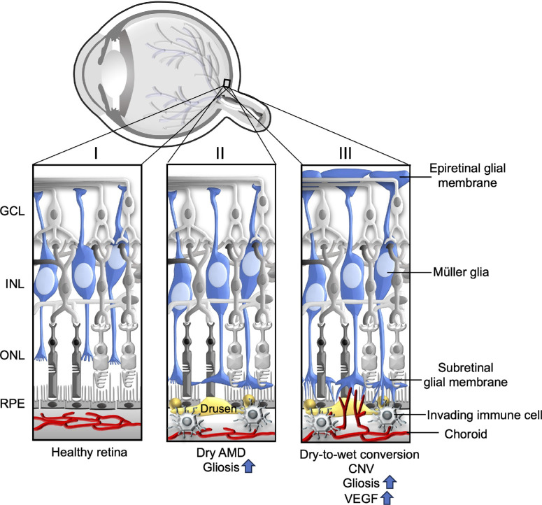
Müller glia, the main glial cell of the retina, are critical for neuronal and vascular homeostasis in the retina. During age-related macular degeneration (AMD) pathogenesis, Müller glial activation, remodeling, and migrations are reported in the areas of retinal pigment epithelial (RPE) degeneration, photoreceptor loss, and choroidal neovascularization (CNV) lesions. Despite this evidence indicating glial activation localized to the regions of AMD pathogenesis, it is unclear whether these glial responses contribute to AMD pathology or occur merely as a bystander effect. In this review, we summarize how Müller glia are affected in AMD retinas and share a prospect on how Müller glial stress might directly contribute to the pathogenesis of AMD. The goal of this review is to highlight the need for future studies investigating the Müller cell's role in AMD. This may lead to a better understanding of AMD pathology, including the conversion from dry to wet AMD, which has no effective therapy currently and may shed light on drug intolerance and resistance to current treatments.


Conflict of interest statement

Disclosure: S. Navneet, None; K. Wilson, None; B. Rohrer, None

Figures

Figure.
Figure.
Schematic representation of the Müller glial response in the healthy, dry, and late-stage or wet AMD retinas. (I) In healthy retinas, Müller glia span the entire thickness of the retina, forming the inner and outer limiting membranes. (II) In dry AMD retinas, activated Müller glia migrate to the outer retina, forming subretinal glial membranes; these GFAP and vimentin-positive glial membranes are explicitly found in the areas of RPE atrophy and choroidal vessel dropout. (III) In retinas with CNV or late-stage AMD, increased epiretinal glial membranes are reported in addition to lesion-specific activated subretinal Müller glial scars. These areas are also rich in recruited immune cells and microglia. GCL, ganglion cell layer; INL, inner nuclear layer; ONL, outer nuclear layer; RPE, retinal pigment epithelium.

References

    1. Bowes Rickman C, Farsiu S, Toth CA, Klingeborn M.. Dry age-related macular degeneration: mechanisms, therapeutic targets, and imaging. Invest Ophthalmol Vis Sci. 2013; 54: Orsf68–Orsf80. - PMC - PubMed
    1. Boyer DS, Schmidt-Erfurth U, van Lookeren Campagne M, Henry EC, Brittain C.. The pathophysiology of geographic atrophy secondary to age-related macular degeneration and the complement pathway as a therapeutic target. Retina. 2017; 37: 819–835. - PMC - PubMed
    1. Ambati J, Atkinson JP, Gelfand BD.. Immunology of age-related macular degeneration. Nat Rev Immunol. 2013; 13: 438–451. - PMC - PubMed
    1. Heier JS, Brown DM, Shah SP, et al. .. Intravitreal aflibercept injection vs sham as prophylaxis against conversion to exudative age-related macular degeneration in high-risk eyes: a randomized clinical trial. JAMA Ophthalmol. 2021; 139: 542–547. - PMC - PubMed
    1. Schmidt-Erfurth U, Waldstein SM, Klimscha S, et al. .. Prediction of individual disease conversion in early AMD using artificial intelligence. Invest Ophthalmol Vis Sci. 2018; 59: 3199–3208. - PubMed