. 2024 Mar 5;121(10):e2313540121.
doi: 10.1073/pnas.2313540121. Epub 2024 Feb 28.

Tom20 gates PINK1 activity and mediates its tethering of the TOM and TIM23 translocases upon mitochondrial stress

Affiliations

Tom20 gates PINK1 activity and mediates its tethering of the TOM and TIM23 translocases upon mitochondrial stress

Mohamed A Eldeeb et al. Proc Natl Acad Sci U S A. .

Abstract

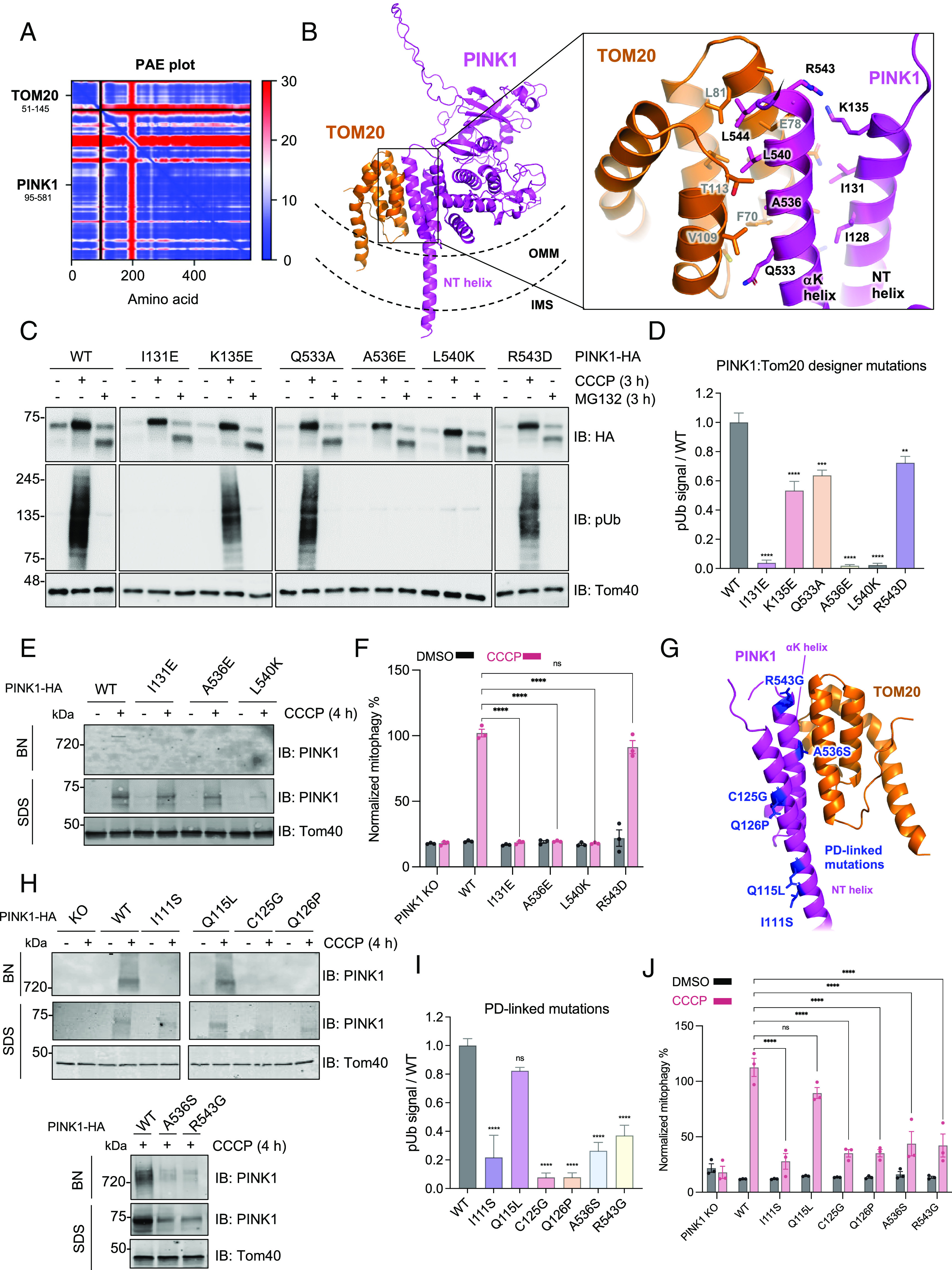
Mutations in PTEN-induced putative kinase 1 (PINK1) cause autosomal recessive early-onset Parkinson's disease (PD). PINK1 is a Ser/Thr kinase that regulates mitochondrial quality control by triggering mitophagy mediated by the ubiquitin (Ub) ligase Parkin. Upon mitochondrial damage, PINK1 accumulates on the outer mitochondrial membrane forming a high-molecular-weight complex with the translocase of the outer membrane (TOM). PINK1 then phosphorylates Ub, which enables recruitment and activation of Parkin followed by autophagic clearance of the damaged mitochondrion. Thus, Parkin-dependent mitophagy hinges on the stable accumulation of PINK1 on the TOM complex. Yet, the mechanism linking mitochondrial stressors to PINK1 accumulation and whether the translocases of the inner membrane (TIMs) are also involved remain unclear. Herein, we demonstrate that mitochondrial stress induces the formation of a PINK1-TOM-TIM23 supercomplex in human cultured cell lines, dopamine neurons, and midbrain organoids. Moreover, we show that PINK1 is required to stably tether the TOM to TIM23 complexes in response to stress such that the supercomplex fails to accumulate in cells lacking PINK1. This tethering is dependent on an interaction between the PINK1 N-terminal-C-terminal extension module and the cytosolic domain of the Tom20 subunit of the TOM complex, the disruption of which, by either designer or PD-associated PINK1 mutations, inhibits downstream mitophagy. Together, the findings provide key insight into how PINK1 interfaces with the mitochondrial import machinery, with important implications for the mechanisms of mitochondrial quality control and PD pathogenesis.

Keywords: PINK1; mitochondrial import; mitochondrial quality control; mitophagy; proteolysis.


Conflict of interest statement

Competing interests statement:J.-F.T. was a member of the scientific board of Mitokinin Inc. at the time of submission. When Mitokinin Inc. was later acquired, he received financial compensation for this consultancy work and is no longer connected with the company.

Figures

Fig. 1.
Fig. 1.
HMW PINK1 complex formation is ubiquitous across cell types and assembles in response to depolarization or misfolded protein accumulation. (A) HEK293T cells (endogenous or expressing PINK1-His6 or PINK1-3×FLAG) were treated with 20 µM CCCP or DMSO for the indicated time points, lysed in 1% digitonin solubilization buffer or sodium dodecyl sulfate (SDS) sample buffer, subjected to BN-PAGE or SDS-PAGE, and immunoblotted. (B) U2OS cells expressing endogenous PINK1 were treated with 20 µM CCCP or for 4 h before washing out CCCP for the indicated times. Cells were lysed and immunoblotted for PINK1 and Tom40 as in A. (C) Schematic of hMBO generation from peripheral blood mononuclear cells which have been reprogrammed into iPSCs and CRISPR edited for PINK1 deletion. (D) Six-week-old PINK1 KO DA neurons were treated with 20 µM CCCP or DMSO, subjected to BN-PAGE or SDS-PAGE, and then immunoblotted. (E) hMBOs (PINK1-KO or WT) that were 8.5 wk old were treated with 20 µM CCCP and 5 mM ammonium chloride to inhibit lysosomal degradation as indicated, subjected to BN-PAGE and SDS-PAGE, and were immunoblotted using PINK1 and Tom40 antibodies. (F) U2OS cells were transiently transfected with vector or ΔOTC constructs for 36 h and then fractionated. Mitochondrial-enriched fractions were solubilized, used for BN-PAGE (Upper) and SDS-PAGE (Lower), and immunoblotted. (G) Quantification of mitophagy using the mt-Keima reporter assay in PINK1-HA transfected U2OS PINK1 KO cells following DMSO (black) or CCCP (violet) treatment, or ΔOTC transfection (pink). Bars indicate the relative level of mitophagy, normalized to WT PINK1 treated with CCCP, plotted as mean (n = 3) ± SEM. Two-way ANOVA with Tukey’s post hoc tests (n = 3), *P < 0.05; **P < 0.01; ***P < 0.001; ****P < 0.0001; ns, not significant. (H) U2OS PINK1 KO cells were transfected PINK1-HA or mock vector and treated with 20 µM CCCP for 4 h. Mitochondria were isolated, and HA immunocapture was performed. Bound proteins were eluted with HA peptide, and fractions were subjected to SDS-PAGE or BN-PAGE immunoblotting. (I) Mock or PINK1-HA transfected U2OS PINK1 KO cells were transfected with ΔOTC for 36 h followed by mitochondrial isolation and immunocapture. Bound proteins were eluted and subjected to SDS-PAGE immunoblotting for the antibodies indicated.
Fig. 2.
Fig. 2.
PINK1 forms a supercomplex with TOM and TIM23 complexes upon depolarization. (A) HEK293T cells were transfected with PINK1-Strep or mock vector, mitochondria were isolated using nitrogen cavitation, StrepTactin purification was performed, and eluates were subjected to mass spectrometry analysis (Left). LFQ was performed (n = 3) and results are plotted as a volcano plot. Significantly enriched hits (−log10 P-value > 1.3 and log2 fold change > 2, indicated by dashed lines) are colored according to their average intensity-based absolute quantification (iBAQ) intensity across PINK1-Strep samples and selected hits are labeled. The core human TOM complex (PDB: 7CK6) is depicted as cartoons and labeled for reference. (B) Mock or pCMV(d1) PINK1-HA transfected U2OS PINK1 KO cells were treated with 20 µM CCCP or DMSO as indicated, subjected to BN-PAGE or SDS-PAGE, and immunoblotted as indicated. (C) Mock or Tim50-FLAG transfected HEK293T cells were treated with 20 µM CCCP for 4 h followed by mitochondrial isolation and FLAG immunocapture. Bound proteins were eluted with FLAG peptide, and fractions were subjected to SDS-PAGE immunoblotting.
Fig. 3.
Fig. 3.
Characterization of the PINK1-Tom20 binding interface using designer and PD-linked mutations within the PINK1 NT-CTE. (A) AlphaFold multimer v3 was run on the cytosolic domains of PINK1 (a.a. 95 to 581) and Tom20 (a.a. 51 to 145) with 20 recycles and an RMSD tolerance between cycles of 0.5 Å. The top-ranked model was chosen for PAE plot visualization. (B) Structural visualization of the PINK1-Tom20 AlphaFold model using PyMOL. PINK1 and Tom20 are depicted in the context of the OMM and IMS topology. (C) U2OS PINK1 KO cells were transfected with pCMV(d1) PINK1-HA (WT or indicated mutants) and were treated with 20 μM CCCP or 10 µM MG132 for 4 h. Lysates were run on SDS-PAGE and immunoblotted as indicated. (D) Quantification of immunoblots by densitometry for C. Band intensities were normalized relative to the level of Tom40 in the CCCP-treated sample, and the ratio to pSer65 of WT CCCP-treated sample was calculated. Bars represent mean ± SEM (n = 3). One-way ANOVA with Dunnett’s post hoc test was performed (*P < 0.05; **P < 0.01; ***P < 0.001; ****P < 0.0001; ns, not significant). (E) U2OS PINK1 KO cells were transfected with pCMV(d1) PINK1-HA WT or mutants and treated with 20 μM CCCP for 4 h. Mitochondria were extracted, solubilized, and analyzed via BN-PAGE or SDS-PAGE immunoblotting. (F) U2OS cells transfected with pCMV(d1) PINK1 (WT or mutants) were treated with 20 μM CCCP or DMSO for 4 h and mitophagy was quantified using the mt-Keima reporter assays. Bars indicate the relative level of mitophagy, normalized to WT PINK1 treated with CCCP, plotted as mean (n = 3) ± SEM. Two-way ANOVA with Tukey’s post hoc tests (n = 3), *P < 0.05; **P < 0.01; ***P < 0.001; ****P < 0.0001; ns, not significant. (G) PD-linked mutations visualized within the PINK1 NT-CTE and TOM20 AlphaFold model. (H) U2OS PINK1 KO cells were transfected with pCMV(d1) PINK1-HA (WT or indicated mutants) and treated with 20 μM CCCP for 4 h. Mitochondria were extracted, solubilized, and analyzed by immunoblotting. (I) U2OS PINK1 KO cells were transfected with pCMV(d1) PINK1-HA (WT or indicated mutants) and treated with 20 μM CCCP for 4 h. Lysates were immunoblotted and quantified as in D. (J) U2OS PINK1 KO cells were transfected with pCMV(d1) PINK1 mutants and assayed using the mt-Keima reporter assay and quantified as in F.
Fig. 4.
Fig. 4.
PINK1 endogenously tethers TOM and TIM23 following import arrest in a Tom20-dependent manner. (A) U2OS cells (WT or PINK1-KO) were transfected with Tom22-FLAG and treated with 20 µM CCCP or DMSO for 4 h, followed by mitochondrial isolation and FLAG immunocapture. Bound proteins were eluted with FLAG peptides and subjected to SDS-PAGE immunoblotting. (B) U2OS cells (WT or PINK1-KO) were transfected with Tim50-FLAG and treated with 20 µM CCCP or DMSO for 4 h, followed by mitochondrial isolation and FLAG immunocapture as in Fig. 3A. (C) U2OS PINK1 KO cells transfected with pCMV(d1) PINK1-HA (WT or Tom20 binding deficient mutants) were treated with 20 µM CCCP for 4 h followed by mitochondrial isolation and HA immunocapture. Bound proteins were eluted with HA peptides and subjected to SDS-PAGE immunoblotting. (D) U2OS PINK1 KO cells expressing PINK1-HA were transfected with mock vector, CRISPRi sgRNA targeting Tim50, or Tim50-Flag, and treated with 20 µM CCCP or DMSO for 4 h. Lysates were subjected to SDS-PAGE or BN-PAGE immunoblotting as previously described. (E) U2OS PINK1 KO cells expressing PINK1-HA were transfected with mock vector or CRISPRi sgRNA targeting Tim50 and treated with 20 µM CCCP or DMSO for 4 h. Lysates were subjected to SDS-PAGE or BN-PAGE immunoblotting for the antibodies indicated as previously described. (F) Simplified schematic of PINK1 topology within the PINK1-TOM-TIM supercomplex. Accessory TOM subunits are depicted as gray. PINK1 NT and αK helices are labeled along with Tom20 (depicted as “20”). This model of Tom20 binding places the TMD of PINK1 within the Tom40 pore in the OMM.

References

    1. Reich S. G., Savitt J. M., Parkinson’s disease. Med. Clin. North Am. 103, 337–350 (2019). - PubMed
    1. Pickrell A. M., Youle R. J., The roles of PINK1, parkin, and mitochondrial fidelity in Parkinson’s disease. Neuron 85, 257–273 (2015). - PMC - PubMed
    1. Martin I., Dawson V. L., Dawson T. M., Recent advances in the genetics of Parkinson’s disease. Annu. Rev. Genomics Hum. Genet. 12, 301–325 (2011). - PMC - PubMed
    1. Eldeeb M. A., Thomas R. A., Ragheb M. A., Fallahi A., Fon E. A., Mitochondrial quality control in health and in Parkinson’s disease. Physiol. Rev. 102, 1721–1755 (2022). - PubMed
    1. Pickles S., Vigié P., Youle R. J., Mitophagy and quality control mechanisms in mitochondrial maintenance. Curr. Biol. 28, R170–R185 (2018). - PMC - PubMed

MeSH terms