. 2024 Mar-Apr;38(2):531-538.
doi: 10.21873/invivo.13471.

Development of a Simple and Reproducible Cell-derived Orthotopic Xenograft Murine Model for Neuroblastoma

Affiliations

Development of a Simple and Reproducible Cell-derived Orthotopic Xenograft Murine Model for Neuroblastoma

Kathleen Doyle et al. In Vivo. 2024 Mar-Apr.

Abstract

Background/aim: Neuroblastoma is a common childhood cancer with poor survival for children with high-risk disease, and ongoing research to improve outcomes is needed. Patient-derived xenografts (PDX) and genetically engineered mouse models (GEMM) are reliable models for oncologic research; however, they are resource-intensive, expensive, and require significant expertise to develop and maintain. We developed an orthotopic xenograft murine model of neuroblastoma that utilizes cryopreserved banks of human neuroblastoma cell lines, requires minimal equipment, and is easily reproducible.

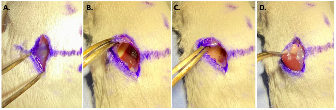
Materials and methods: The neuroblastoma cell line NB1643 was obtained from the Children's Oncology Group (COG) Childhood Cancer Repository. Nod-SCID-gamma (NSG) mice underwent orthotopic injection of 2x10⁶ NB1643 cells suspended in 10 μl of collagen hydrogel directly into the adrenal gland via an open retroperitoneal surgical approach. Mice were monitored by ultrasound and in vivo imaging system (IVIS) until the tumor reached the volume of the ipsilateral kidney. Tumor identity was confirmed by necropsy and histologic analysis.

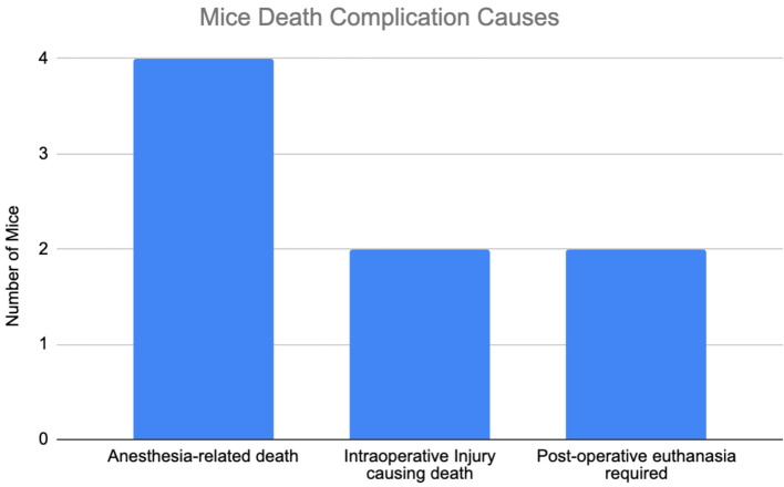
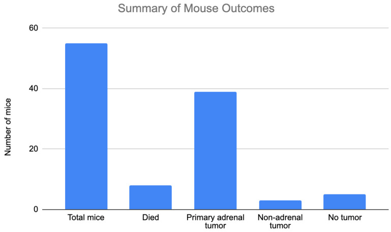
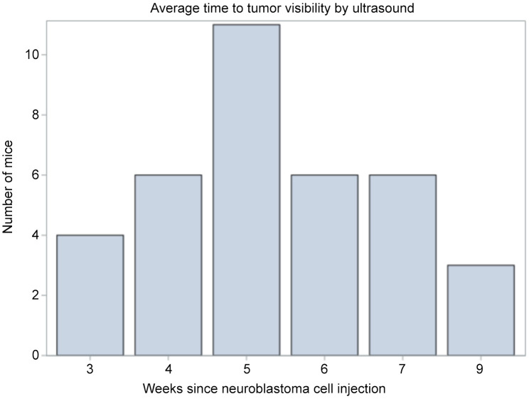
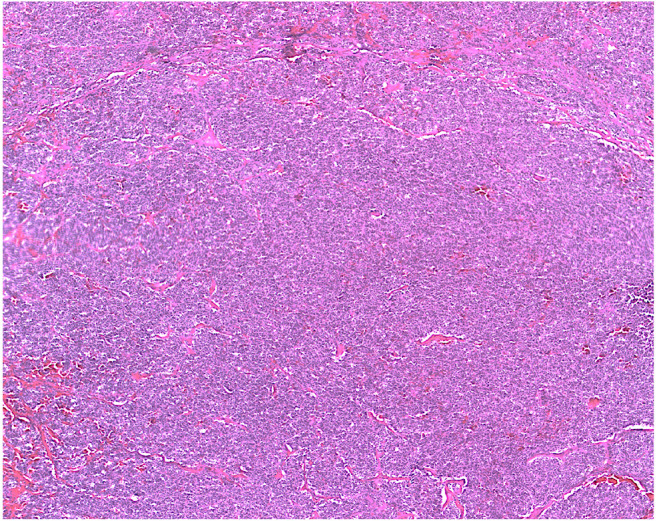
Results: A total of 55 mice underwent surgery. Eight died due to anesthetic or surgical complications. 39/47 (78%) survivors grew primary adrenal tumors. Average anesthesia time was 30 min. Ultrasound and IVIS successfully characterized tumor growth in all mice. Average time to target tumor size was 5 weeks (range=3-9). Gross pathologic and histologic analysis confirmed adrenal tumors consistent with neuroblastoma in all mice with adrenal masses.

Conclusion: A cell-derived orthotopic xenograft murine model can be successfully used to create an in vivo model of neuroblastoma. This model can be utilized in environments where PDX or GEMM models are not feasible.

Keywords: Neuroblastoma; PDOX; orthotopic murine model.


Conflict of interest statement

The Authors have no conflicts of interest to disclose related to the above manuscript.

Figures

Figure 1
Figure 1. Incision location. (A) Mouse spinal column convexity (dotted line) is primary anatomical landmark for incision location. Crossed solid lines represent incision location: 1 cm caudal from the most convex portion of the spinal column and one cm lateral and to the left of the midline. (B) Incision length is 1 cm and made in the cephalad to caudad direction. (C) Incision is 1 cm lateral and to the left to midline.
Figure 2
Figure 2. Surgical technique. (A) The incision was made through the skin. Image demonstrates retroperitoneal fat plane that is encountered after skin incision. Through the retroperitoneal fat the kidney can be visualized to ensure the incision is not too low or too high. (B) After incision through the retroperitoneum the left lobe of the liver is often encountered. (C) The left lobe of the liver should be bluntly and gently retracted cephalad and lateral to expose the kidney, which is shown on the right side of the incision here. (D) the adrenal gland is exposed. The adrenal gland can be gently brought into the incision by grasping the surrounding fat pad to avoid injury to the adrenal gland or kidney.
Figure 3
Figure 3. Injection of neuroblastoma cells. Image showing the syringe containing neuroblastoma cells directly being injected into the left adrenal gland.
Figure 4
Figure 4. In vivo imaging system monitoring of tumor growth. (A) Schematic for mechanism of bioluminescent imaging via IVIS. Neuroblastoma cells (NB1643) were transfected with luciferase gene via plasmid. Mice were injected with luciferin and a bioluminescent signal was emitted and measured by IVIS machine. (B) Serial weekly images demonstrating presence of neuroblastoma cells in the mouse and then coalescence of the cell signal to the left adrenal gland as the tumor grows and develops. IVIS: In vivo imaging system.
Figure 5
Figure 5. Tumor image on ultrasound. Five weeks post NB cell injection. Diameter: 0.79 cm and area: 0.40 cm2. NB: Neuroblastoma.
Figure 6
Figure 6. Complications leading to murine death prior to tumor development. Four mice died from anesthetic complications during surgery or postoperative imaging. Two mice had intraoperative injuries that resulted in death. Two mice required post-operative euthanasia for reasons unrelated to surgery (one mouse had a self-inflicted tail injury requiring euthanasia and one mouse was found to have been injected with neuroblastoma cells contaminated with mycoplasma).
Figure 7
Figure 7. Summary of mouse outcomes. A total of 55 mice underwent surgery. Eight died due to surgical or anesthetic complications. Thirty-nine mice grew primary adrenal tumors resulting in an engraftment rate of 83%. Three mice had non-adrenal lesions. Five mice had no tumor growth.
Figure 8
Figure 8. Weeks to tumor visibility on ultrasound from time of neuroblastoma cell injection. Average time was 5 weeks with a range of 3-9 weeks.
Figure 9
Figure 9. Histology results. Hematoxylin and eosin (H&E) staining of adrenal tumor (20×). Sample demonstrates small, round, blue cells consistent with neuroblastoma.

References

    1. Hochheuser C, Windt LJ, Kunze NY, de Vos DL, Tytgat GAM, Voermans C, Timmerman I. Mesenchymal stromal cells in neuroblastoma: exploring crosstalk and therapeutic implications. Stem Cells Dev. 2021;30(2):59–78. doi: 10.1089/scd.2020.0142. - DOI - PMC - PubMed
    1. Davidoff AM. Neuroblastoma. Semin Pediatr Surg. 2012;21(1):2–14. doi: 10.1053/j.sempedsurg.2011.10.009. - DOI - PMC - PubMed
    1. Upton K, Modi A, Patel K, Kendsersky NM, Conkrite KL, Sussman RT, Way GP, Adams RN, Sacks GI, Fortina P, Diskin SJ, Maris JM, Rokita JL. Epigenomic profiling of neuroblastoma cell lines. Sci Data. 2020;7(1):116. doi: 10.1038/s41597-020-0458-y. - DOI - PMC - PubMed
    1. VAN Noord RA, Thomas T, Krook M, Chukkapalli S, Hoenerhoff MJ, Dillman JR, Lawlor ER, Opipari VP, Newman EA. Tissue-directed implantation using ultrasound visualization for development of biologically relevant metastatic tumor xenografts. In Vivo. 2017;31(5):779–791. doi: 10.21873/invivo.11131. - DOI - PMC - PubMed
    1. Hidalgo M, Amant F, Biankin AV, Budinská E, Byrne AT, Caldas C, Clarke RB, de Jong S, Jonkers J, Mælandsmo GM, Roman-Roman S, Seoane J, Trusolino L, Villanueva A. Patient-derived xenograft models: an emerging platform for translational cancer research. Cancer Discov. 2014;4(9):998–1013. doi: 10.1158/2159-8290.CD-14-0001. - DOI - PMC - PubMed