. 2024 Feb 28;15(1):1831.
doi: 10.1038/s41467-024-46096-z.

Cryo-EM structure of cell-free synthesized human histamine 2 receptor/Gs complex in nanodisc environment

Affiliations

Cryo-EM structure of cell-free synthesized human histamine 2 receptor/Gs complex in nanodisc environment

Zoe Köck et al. Nat Commun. .

Abstract

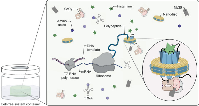
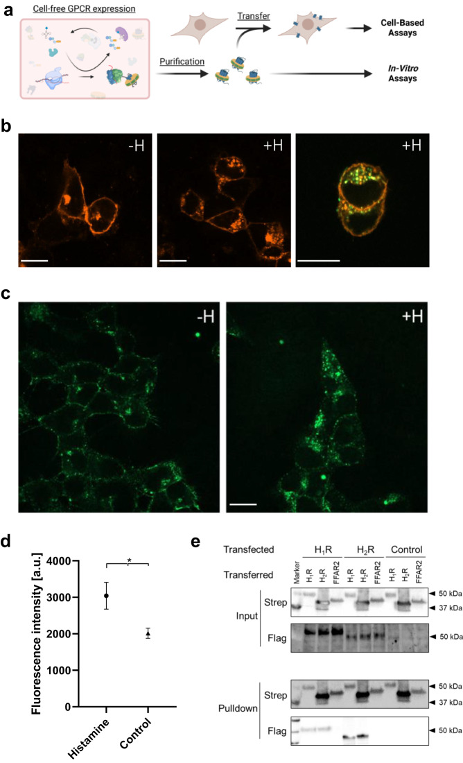
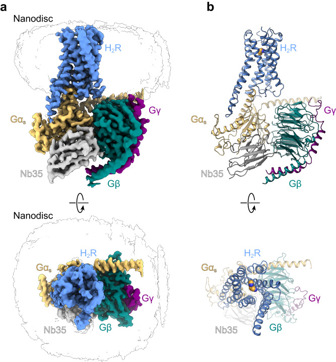
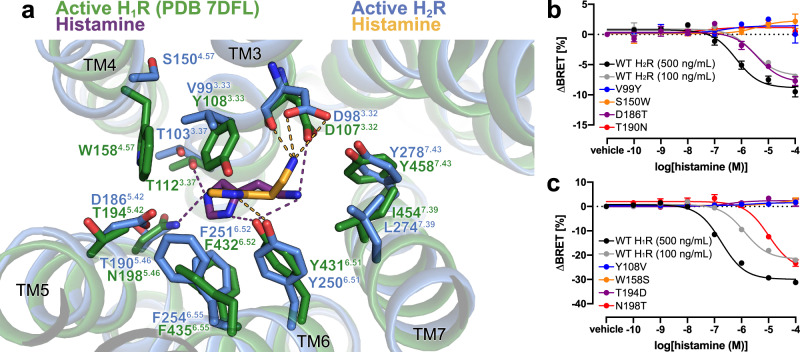
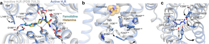
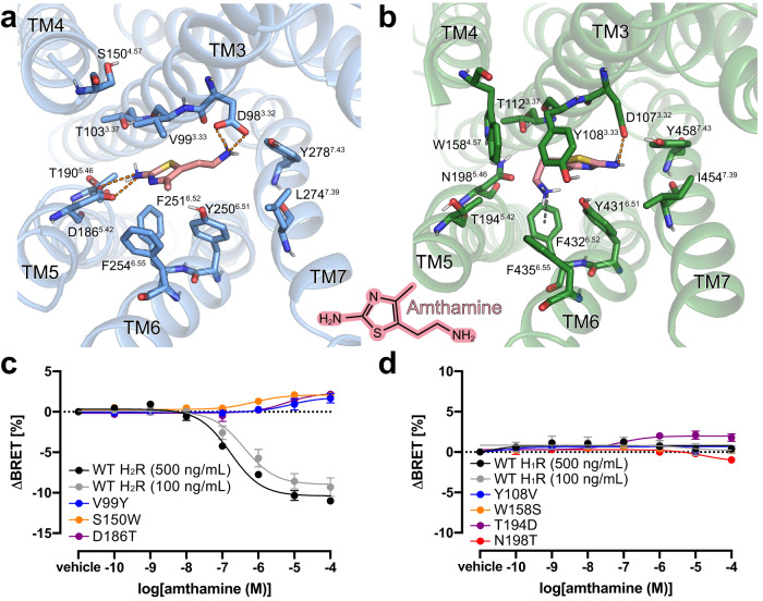
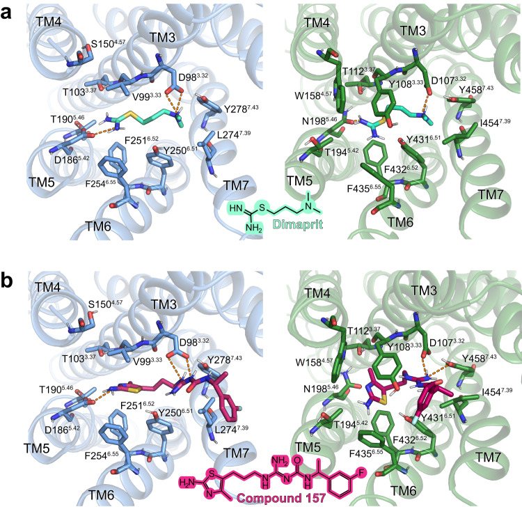
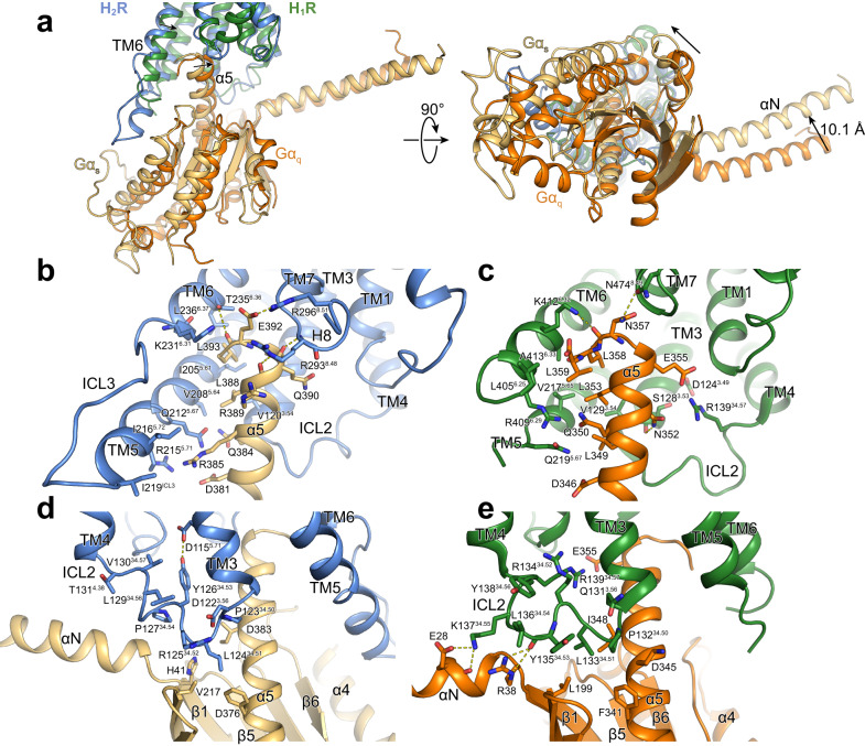
Here we describe the cryo-electron microscopy structure of the human histamine 2 receptor (H2R) in an active conformation with bound histamine and in complex with Gs heterotrimeric protein at an overall resolution of 3.4 Å. The complex was generated by cotranslational insertion of the receptor into preformed nanodisc membranes using cell-free synthesis in E. coli lysates. Structural comparison with the inactive conformation of H2R and the inactive and Gq-coupled active state of H1R together with structure-guided functional experiments reveal molecular insights into the specificity of ligand binding and G protein coupling for this receptor family. We demonstrate lipid-modulated folding of cell-free synthesized H2R, its agonist-dependent internalization and its interaction with endogenously synthesized H1R and H2R in HEK293 cells by applying a recently developed nanotransfer technique.


Conflict of interest statement

The authors declare no competing interests.

Figures

Fig. 1
Fig. 1. Cryo-EM sample preparation of complexes by CF expression.
H2R is synthesized by cotranslational insertion into preformed NDs (DOPG) in the presence of histamine and Gs heterotrimer purified from insect cells (Created with BioRender.com).
Fig. 2
Fig. 2. Characterization of CF synthesized H2R after nanotransfer into HEK293 cells.
a Schematic of GPCR nanotransfer. b H1R-mCherry synthesized after transfection internalizes upon histamine treatment (100 µM histamine for 1 h; +H) and co-localizes with nanotransferred and CF synthesized H2R-mNG (right panel). White bar = 10 µm. Representative images from three independent experiments. c HEK293 cells transferred for 4 h with 0.5 µM purified H2R-mNG in NDs (DOPG) and treated with histamine. Left panel: Localization of transferred H2R-mNG in untreated cells. Right panel: Internalization of transferred H2R after incubation with 100 µM histamine for 1 h. White bar = 10 µm. Representative images from three independent experiments. d Quantification of cytosolic fluorescence of transferred H2R-mNG. Data are presented as mean (SD) (n = 3 individual experiments with ≥ six data points each, *P < 0.05, two-sided students t-test). Source data are provided as Source Data file. e Interaction of transferred CF synthesized GPCRs with transfected H1R and H2R. Transfected cells expressing Flag-tagged H1R or H2R were transferred with CF synthesized Strep-tagged H1R, H2R, or FFAR2 for 4 h. Subsequently, cells were washed, lysed and transferred GPCRs were immobilized by anti-Strep pulldowns. Input: Immunoblots of cell lysates before anti-Strep pulldowns. Pulldown: Immunoblots of immobilized GPCRs after anti-Strep pulldowns. Strep: Anti-Strep antibody; Flag: Anti-Flag antibody; Control: Cells without transfection. The experiment was performed once, uncropped figures are provided in the Source Data File. (Fig. 2a, created with BioRender.com).
Fig. 3
Fig. 3. Cryo-EM structure of the histamine-bound H2R-Gs/Nb35/ND complex.
a Cryo-EM density map of the histamine-bound H2R/Gs/Nb35/ND complex colored by subunit. Blue, H2R; wheat, Gαs; cyan, Gβ; purple, Gγ, grey, Nb35; black silhouettes, ND. b Ribbon model of the H2R/Gs/Nb35/ND complex bound to histamine (yellow spheres) colored by subunit.
Fig. 4
Fig. 4. Histamine-binding to H2R.
a Comparison of the histamine-binding site in H2R (blue, receptor; yellow, histamine) and H1R (green, receptor; purple, histamine) (PDB ID 7DFL). Binding pocket residues are represented as sticks and labeled with residue number and Ballesteros-Weinstein code (superscript) colored by the receptor subtype. H-bond interactions are shown as dotted lines colored by the interacting ligand. b, c Mutagenesis analysis of receptor-specific residues within the histamine-binding pocket in H2R b and H1R c using a BRET-based G protein dissociation assay. Values in brackets represent the amount of DNA (ng) of receptor plasmid used to transfect one mL of cells. 500 ng DNA per mL of cells was used for all mutants. Signaling graphs show mean ± s.e.m. of three independent biological replicates with a global fit of the data. Source data are provided as Source Data file.
Fig. 5
Fig. 5. H2R activation by histamine.
a Binding pocket of the histamine-activated H2R (blue) with the inactive famotidine-bound receptor (gray) (PDB ID 7UL3) overlaid. H-bond interactions are shown as dotted lines colored by the interacting ligand. b Close-up view of the CWxP and PIF motif in the inactive (grey) H2R and the active (blue) H2R bound to histamine (yellow spheres). c Comparison of the DRY motif and the NPxxY motif in the inactive (grey) H2R and active (blue) H2R structures. Amino acid side chains are represented as sticks and labeled with residue number and Ballesteros-Weinstein code (superscript) colored by the receptor subtype. Arrows indicate conformational changes in the H2R upon activation.
Fig. 6
Fig. 6. Agonist binding selectivity of H2R and H1R.
a and b Predicted binding modes for the H2R selective agonist amthamine (light pink) in a H2R (blue) and b H1R (green). The orange dotted lines represent H-bond interactions. The grey dotted line represents a possible cation-aromatic interaction between F4326.52 in H1R and the protonated amino group of amthamine. Binding pocket residues are represented as sticks and labeled with residue number and Ballesteros-Weinstein code (superscript). Binding mode predictions were obtained with AutoDock Vina. c, d Mutagenesis analysis of receptor-specific residues within the ligand-binding pocket in c H2R and d H1R using a BRET-based G protein dissociation assay in the presence of amthamine. Values in brackets represent the amount of DNA (ng) of receptor plasmid used to transfect one mL of cells. 500 ng DNA per mL of cells was used for all mutants. Signaling graphs show mean ± s.e.m. of three independent biological replicates with a global fit of the data.
Fig. 7
Fig. 7. Molecular docking of H2R specific agonists in the H2R and the H1R.
a Docking of dimaprit; b Docking of compound 157. Structural elements and relevant residues of the binding pockets of the H2R (blue) or the H1R (green) are shown. The orange dotted lines represent H-bond interactions. Binding pocket residues are represented as sticks and labeled with residue number and Ballesteros-Weinstein number (superscript). Binding mode predictions were obtained with AutoDock Vina.
Fig. 8
Fig. 8. Comparison of G protein interfaces between the H2R-Gs and the H1R-Gq complex.
a Comparison of the relative receptor-G protein orientation between the H2R (blue) and the H1R (green) subtype coupled to Gαs (wheat) and Gαq (orange), respectively. b, c Comparison of the interface between b the α5 of Gαs and H2R and c the α5 of Gαq and H1R. d, e Comparison of the interface between the ICL2 and the G protein subtype in d the H2R-Gαs complex and e the H1R-Gαq complex. Interface residues are represented as sticks and labeled with residue number and for the receptor residues with the Ballesteros-Weinstein code (superscript). Arrows indicate differences in the relative orientation of the G protein subtypes with respect to the receptors.

Similar articles

Cited by

References

    1. Parsons ME, Ganellin CR. Histamine and its receptors. Br. J. Pharmacol. 2006;147:127–135. doi: 10.1038/sj.bjp.0706440. - DOI - PMC - PubMed
    1. Jorgensen EA, Knigge U, Warberg J, Kjaer A. Histamine and the regulation of body weight. Neuroendocrinology. 2007;86:210–214. doi: 10.1159/000108341. - DOI - PubMed
    1. Jutel M, Akdis M, Akdis CA. Histamine, histamine receptors and their role in immune pathology. Clin. Exp. Allergy. 2009;39:1786–1800. doi: 10.1111/j.1365-2222.2009.03374.x. - DOI - PubMed
    1. Delvalle J, Wang L, Gantz I, Yamada T. Characterization of H2 histamine receptor: linkage to both adenylate cyclase and [Ca2+]i signaling systems. Am. J. Physiol. -Gastr. L. 1992;263:G967–G972. - PubMed
    1. Panula P, et al. International union of basic and clinical pharmacology. XCVIII. Histamine receptors. Pharmacol. Rev. 2015;67:601–655. doi: 10.1124/pr.114.010249. - DOI - PMC - PubMed