. 2024 Feb 28;15(1):1835.
doi: 10.1038/s41467-024-46047-8.

BTLA contributes to acute-on-chronic liver failure infection and mortality through CD4+ T-cell exhaustion

Affiliations

BTLA contributes to acute-on-chronic liver failure infection and mortality through CD4+ T-cell exhaustion

Xueping Yu et al. Nat Commun. .

Abstract

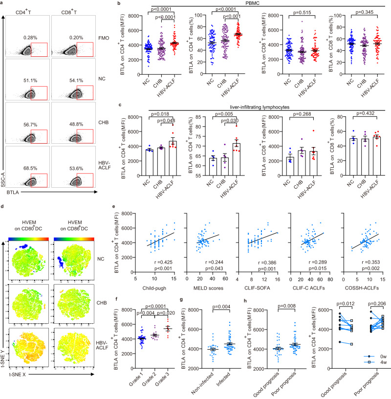
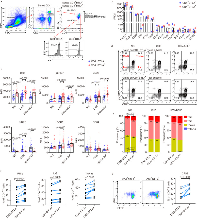
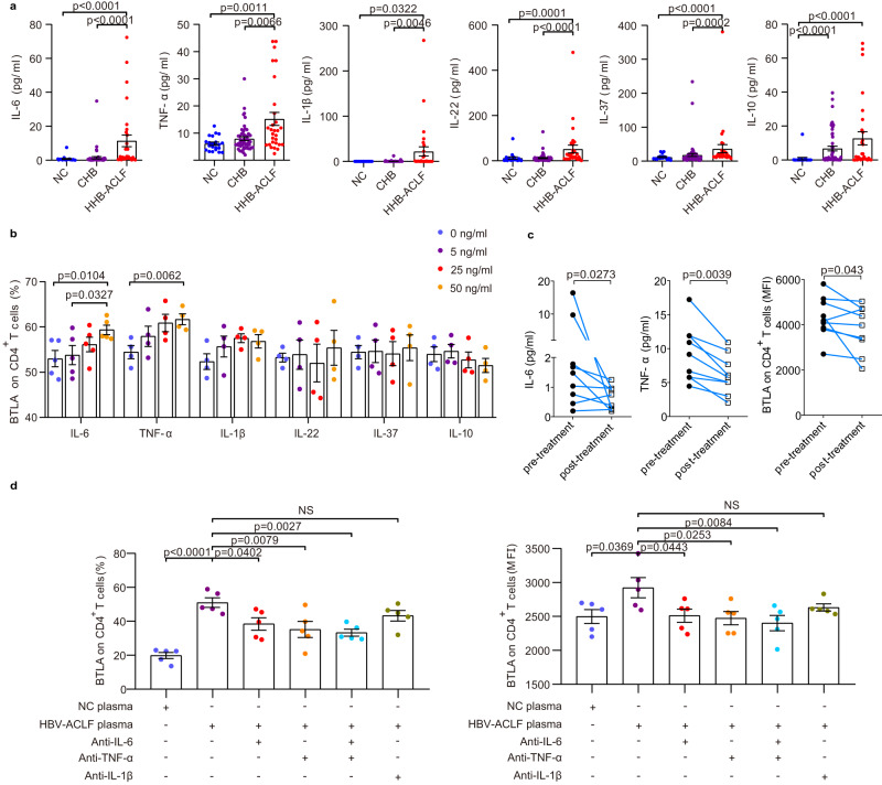
B- and T-lymphocyte attenuator (BTLA) levels are increased in patients with hepatitis B virus-related acute-on-chronic liver failure (HBV-ACLF). This condition is characterized by susceptibility to infection and T-cell immune exhaustion. However, whether BTLA can induce T-cell immune exhaustion and increase the risk of infection remains unclear. Here, we report that BTLA levels are significantly increased in the circulating and intrahepatic CD4+ T cells from patients with HBV-ACLF, and are positively correlated with disease severity, prognosis, and infection complications. BTLA levels were upregulated by the IL-6 and TNF signaling pathways. Antibody crosslinking of BTLA activated the PI3K-Akt pathway to inhibit the activation, proliferation, and cytokine production of CD4+ T cells while promoting their apoptosis. In contrast, BTLA knockdown promoted their activation and proliferation. BTLA-/- ACLF mice exhibited increased cytokine secretion, and reduced mortality and bacterial burden. The administration of a neutralizing anti-BTLA antibody reduced Klebsiella pneumoniae load and mortality in mice with ACLF. These data may help elucidate HBV-ACLF pathogenesis and aid in identifying novel drug targets.


Conflict of interest statement

The authors declare no competing interests.

Figures

Fig. 1
Fig. 1. BTLA expression was significantly increased on circulating and intrahepatic CD4+ T cells in patients with HBV-ACLF and was positively correlated with prognosis and infectious complications.
a Flow cytometry diagram of BTLA expression on peripheral blood CD4+ T and CD8+ T cells in NC, CHB, and HBV-ACLF patients. Patients with HBV-ACLF had significantly increased expression of BTLA on peripheral blood CD4+ T cells (b, NC: n = 90 donors, CHB: n = 104 donors, HBV-ACLF: n = 71 donors) and intrahepatic CD4+ T cells (c, NC: n = 5 donors, CHB: n = 5 donors, HBV-ACLF: n = 7 donors) compared to NC and CHB patients, while there was no significant difference in the BTLA expression on CD8+ T cells from PBMC or from liver-infiltrating lymphocytes. d T-distributed stochastic neighbor embedding (t-SNE) was used to illustrate the increased expression of HVEM on CD80/CD86+ dendritic cells in patients with HBV-ACLF. e The mean fluorescence intensity (MFI) of BTLA expression on CD4+ T cells was positively correlated with the severity of the disease (Child-Pugh, MELD scores, CLIF-SOFA, CLIF-C ACLFs, and COSSH-ACLFs, n = 71 donors). f Changes in the MFI of CD4+BTLA+ T cells in the progression of HBV-ACLF (n = 71 donors). The MFI of CD4+BTLA+ T cells was increased in the coinfection patients (g, n = 44 donors) and poor prognosis group (h, left, n = 39 donors) compared with that in non-infected patients (n = 27 donors) and in those with good prognosis (n = 32 donors), respectively. h right HBV-ACLF patients with good prognosis (n = 9 donors), but not those with poor prognosis (n = 11 donors), had a significantly decreased MFI of CD4+BTLA+ T cells after 4 weeks of treatment. Data were calculated as mean ± SEM (b, c, f, g, h), Kruskal-Wallis H test followed by Dunn’s multiple comparison test (b, c, f), Mann–Whitney U test (g, h left) and Wilcoxon test (h, right). A two-sided P < 0.05 was considered significant.
Fig. 2
Fig. 2. Phenotypic characteristics of BTLA+CD4+ T cells.
a Flow cytometry was used to sort different BTLA subpopulations, followed by RNA-seq. b Differently expressed genes (DEGs) between BTLA+CD4+ T-cell subsets and BTLA-CD4+ T-cell subsets (all n = 2 donors). c Validation of DEGs in BTLA+CD4+ T-cell subsets and BTLA-CD4+ T-cell subsets in NC (n = 38 donors), CHB (n = 94 donors) and HBV-ACLF patients (n = 35 donors). d Representative flow cytometry of BTLA+CD4+ T-cell subsets and BTLA-CD4+ T-cell subsets. e Differentiation phenotypes of BTLA+ CD4+ T cells in NC (n = 26 donors), CHB (n = 17 donors), and HBV-ACLF patients (n = 13 donors). Compared with BTLA-CD4+T cells, BTLA+CD4+T cells secreted higher levels of IFN-γ, IL-2, and TNF-α (f, n = 10 donors), and showed a greater proliferation ability (g, n = 10 donors). Data were calculated as mean ± SEM (c), Two-way ANOVA followed by Sidak’s multiple-comparison test (c), Mann–Whitney test (e), and Wilcoxon test (f, g). A two-sided P < 0.05 was considered significant.
Fig. 3
Fig. 3. Elevated BTLA expression on CD4+ T cells was induced by circulating inflammation cytokines in HBV-ACLF.
a The levels of pro-inflammatory cytokines (IL-1β, IL-6, IL-22, and TNF-α) and anti-inflammatory cytokines (IL-10 and IL-37) were markedly increased in HBV-ACLF patients (n = 29 donors) compared with those in NC (n = 21 donors) and CHB patients (n = 53 donors). b Only rhIL-6 and rhTNF-α significantly increased BTLA expression on CD4+ T cells in a dose-dependent manner (n = 4 donors). c Comprehensive treatment (n = 9 donors) decreased the levels of IL-6 and TNF-α, as well as BTLA expression on CD4+ T cells. d Exposure of purified CD4+ T cells from NC to HBV-ACLF plasma (n = 13 donors) resulted in a higher BTLA expression on CD4+ T cells compared with exposure of PBMC to NC plasma, while HBV-ACLF plasma combined with anti-IL-6 and/or anti-TNF-α resulted in decreased BTLA expression on CD4+ T cells. Data were calculated as mean ± SEM (a, b, d), Kruskal-Wallis H test followed by Dunn’s multiple comparison test (a), Wilcoxon test (c), and one-way ANOVA followed by Tukey’s (b) or Dunnett’s multiple comparison test (d). A two-sided P < 0.05 was considered significant.
Fig. 4
Fig. 4. Crosslinked BTLA markedly inhibited the activation, production of cytokines, and proliferation of BTLA+CD4+ T cells but promoted apoptosis.
a Flow cytometry for CD25, CD38, and CD69 (n = 8 donors), (b) production of IFN-γ, IL-2, and TNF-α induced by phorbol 12-myristate 13-acetate /ionomycin (n = 12 donors), (c, d) proliferation (n = 8 donors), and apoptosis of BTLA+CD4+ T cells from NC (n = 8 donors) in the control and crosslinked anti-BTLA groups. Wilcoxon test (ad). A two-sided P < 0.05 was considered significant.
Fig. 5
Fig. 5. BTLA knockdown increased activation, production of cytokines, and proliferation of CD4+ T cells but abrogated apoptosis.
a BTLA expression (left) and MFI (right) in freshly isolated PBMC activated for 3 days in the presence of control shRNA and varying concentrations of BTLA shRNA (n = 5 donors). b Carboxyfluorescein succinimidyl ester (CFSE) indicates the proliferation of the CD4+ T cells that were activated by anti-CD3/CD28 stimulation for 5 days in the presence of control shRNA and BTLA shRNA (left). The quantification (right) is presented as a division index (n = 6 donors). c Contour plots (up) and line graphs (down) display frequencies of Annexin V+CD4+ T cells in the presence of control shRNA and BTLA shRNA (n = 6 donors). d Contour plots (left) and line graphs (right) display frequencies of IFN-γ, IL-2, and TNF-α-expressing cells on CD4+ T cells induced by PMA/ionomycin for 6 h in the presence of control shRNA and BTLA shRNA (n = 6 donors). e Expression of CD25, CD38, and CD69 on CD4+ T cells activated by anti-CD3/CD28 for 1 day in the presence of control shRNA and BTLA shRNA. All PBMC were isolated from NC (n = 6 donors). Data were calculated as mean ± SEM (a), one-way ANOVA followed by Dunnett’s multiple comparison test (a), and Wilcoxon test (be). A two-sided P < 0.05 was considered significant.
Fig. 6
Fig. 6. Crosslinking of BTLA phosphorylated the SHP1/2 protein and activated the PI3K-Akt-GSK-3β pathway.
a, b Venn diagram (left) and heatmap (right) of DEGs, 237 of which were shared by NC (n = 3 donors), CHB (n = 3 donors) and HBV-ACLF patients (n = 3 donors). c KEGG pathway analysis showed that the PI3K-Akt pathway was significantly different, with the largest DEG in the upregulated signaling pathways. d Crosslinking of BTLA phosphorylates the SHP1/2 protein and activates the PI3K-Akt-GSK-3β pathway in purified CD4+ T cells. Data represent two experiments.
Fig. 7
Fig. 7. BTLA expression contributed to immune exhaustion, increased mortality rate, and bacterial burden in the ACLF model induced by ConA.
a, b Schematic diagram (left) of ACLF induced by continuous injection of ConA (8 mg/kg) and hematoxylin-eosin diagrams (right) of liver pathology at baseline and days 8 and 14 in WT (n = 10 mice) and BTLA-/- (n = 10 mice) C57BL/6 mice. c BTLA expression on CD4+ T cells was significantly increased on days 8 and 14, compared to baseline, in WT mice (n = 10 mice) following ConA injection. d The percentages and MFI of activation indices (CD25, CD38, and CD69) were higher in BTLA-/- mice (n = 10 mice) than in WT mice (n = 10 mice) following ConA injection on day 14. e The levels of IFN-γ and TNF-α were higher in BTLA-/- mice (n = 10 mice) than in WT mice (n = 10 mice) at days 8 and (or) 14. f The cecal ligation and puncture (CLP) model of sepsis was established based on WT (n = 8 mice) and BTLA-/- ACLF mice (n = 9 mice) induced by continuous ConA injection. g Following CLP, BTLA-/- mice were significantly protected from mortality compared to WT mice. h Bacterial DNA levels in whole blood were significantly decreased in BTLA-/- ACLF mice (n = 9 mice) compared to those in WT mice (n = 8 mice) following CLP. Data were calculated as mean ± SEM (ce), one-way ANOVA followed by Dunnett’s multiple comparison test (c), Two-way ANOVA followed by Sidak’s multiple-comparison test (d, e), Wilcoxon test (h), log-rank test for survival study (g). A two-sided P < 0.05 was considered significant.
Fig. 8
Fig. 8. BTLA contributed to immune exhaustion and increased mortality and infection rates in the ACLF model induced by CCl4.
a Schematic diagram of ACLF induced by continuous injection of CCl4 (0.2 mL/kg, twice a week) for 8 weeks and subsequent injection of a double dose of CCl4 (0.4 mL/kg). b Hematoxylin-eosin diagram of liver pathology at baseline, 8 weeks, and 8 week + 3 days in WT (n = 10 mice) and BTLA-/- (n = 10 mice) C57BL/6 mice. c Cytokine (TNF-α, IL-2, and IFN-γ) production in CD4+ T cells was higher in BTLA-/- mice (n = 10 mice) than in WT mice (n = 10 mice) at 6 and 8 weeks. d Cytokine (TNF-α, IL-2, and IFN-γ) levels were markedly increased in the plasma of BTLA-/- mice (n = 10 mice) compared to those in WT mice (n = 10 mice) after a double dose of CCl4 injection. e, g Schematic diagram of intraperitoneal (i.p.) K.P. and anti-BTLA antibodies. f, h Survival rate and K.P. load in peripheral blood of both BTLA-/- (n = 10 mice) and WT mice injected with anti-BTLA antibodies (n = 10 mice) were higher than those in the WT (n = 10 mice) and anti-IgG injection groups (n = 9 mice). Data were calculated as mean ± SEM (c, d, f, h), two-way ANOVA followed by Sidak’s multiple-comparison test (c, d), Wilcoxon test (f, h right), log-rank test for survival study (f, h left). A two-sided P < 0.05 was considered significant.

Similar articles

Cited by

References

    1. Arroyo V, Moreau R, Jalan R, Gines P, Study E-CCC. Acute-on-chronic liver failure: a new syndrome that will re-classify cirrhosis. J. Hepatol. 2015;62:S131–S143. doi: 10.1016/j.jhep.2014.11.045. - DOI - PubMed
    1. Victor Dong CJK. Acute-on-chronic liver failure: objective admission and support criteria in the intensive care unit. JHEP Rep. 2019;1:44–52. doi: 10.1016/j.jhepr.2019.02.005. - DOI - PMC - PubMed
    1. Cai J, Zhang M, Han T, Jiang HQ. Characteristics of infection and its impact on short-term outcome in patients with acute-on-chronic liver failure. Medicine. 2017;96:e8057. doi: 10.1097/MD.0000000000008057. - DOI - PMC - PubMed
    1. Fernandez J, et al. Bacterial and fungal infections in acute-on-chronic liver failure: prevalence, characteristics and impact on prognosis. Gut. 2018;67:1870–1880. doi: 10.1136/gutjnl-2017-314240. - DOI - PubMed
    1. Martin-Mateos R, Alvarez-Mon M, Albillos A. Dysfunctional Immune Response in acute-on-chronic liver failure: it takes two to tango. Front. Immunol. 2019;10:973. doi: 10.3389/fimmu.2019.00973. - DOI - PMC - PubMed