

Understanding activity-stability tradeoffs in biocatalysts by enzyme proximity sequencing
- PMID: 38418512
- PMCID: PMC10902396
- DOI: 10.1038/s41467-024-45630-3
Understanding activity-stability tradeoffs in biocatalysts by enzyme proximity sequencing
Abstract
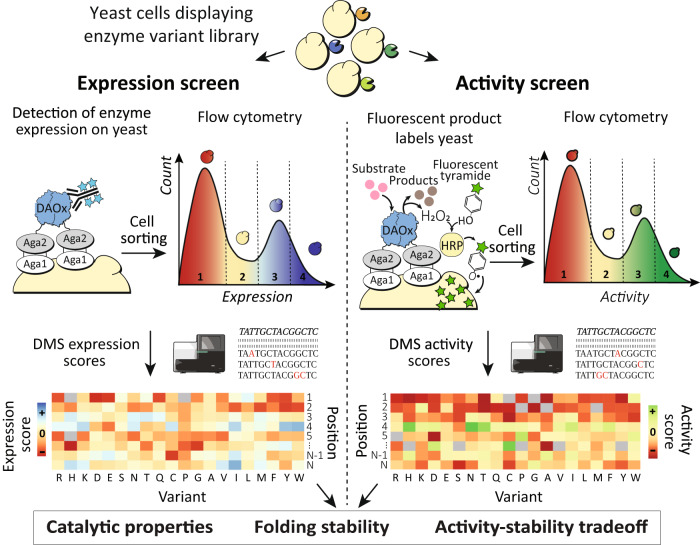
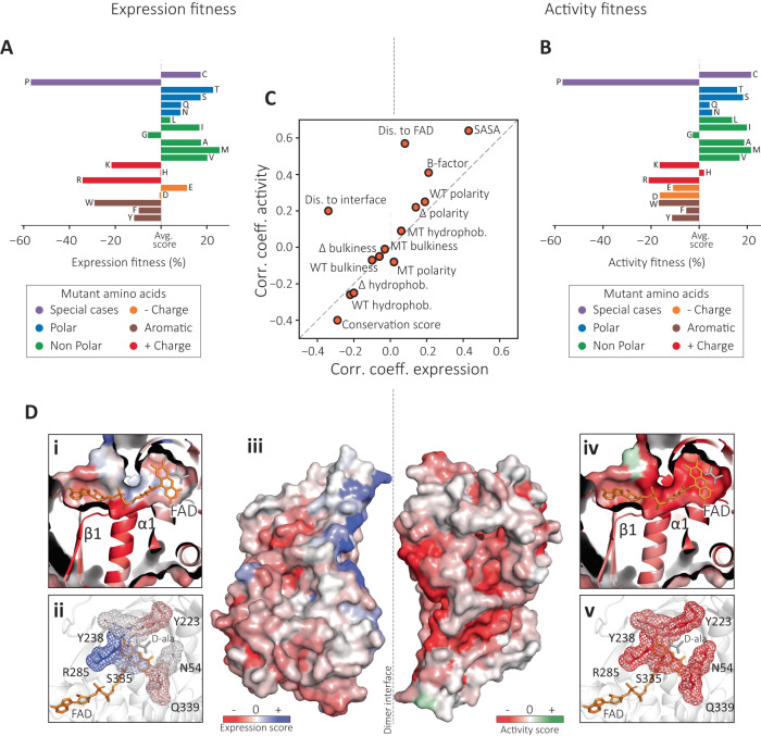
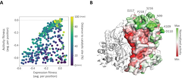
Understanding the complex relationships between enzyme sequence, folding stability and catalytic activity is crucial for applications in industry and biomedicine. However, current enzyme assay technologies are limited by an inability to simultaneously resolve both stability and activity phenotypes and to couple these to gene sequences at large scale. Here we present the development of enzyme proximity sequencing, a deep mutational scanning method that leverages peroxidase-mediated radical labeling with single cell fidelity to dissect the effects of thousands of mutations on stability and catalytic activity of oxidoreductase enzymes in a single experiment. We use enzyme proximity sequencing to analyze how 6399 missense mutations influence folding stability and catalytic activity in a D-amino acid oxidase from Rhodotorula gracilis. The resulting datasets demonstrate activity-based constraints that limit folding stability during natural evolution, and identify hotspots distant from the active site as candidates for mutations that improve catalytic activity without sacrificing stability. Enzyme proximity sequencing can be extended to other enzyme classes and provides valuable insights into biophysical principles governing enzyme structure and function.
© 2024. The Author(s).
Conflict of interest statement
The authors declare no competing interests.
Figures


Similar articles
-
Facile Method for High-throughput Identification of Stabilizing Mutations.J Mol Biol. 2023 Sep 15;435(18):168209. doi: 10.1016/j.jmb.2023.168209. Epub 2023 Jul 20. J Mol Biol. 2023. PMID: 37479080
-
Compensatory stabilizing role of surface mutations during the directed evolution of dienelactone hydrolase for enhanced activity.Protein J. 2015 Feb;34(1):82-9. doi: 10.1007/s10930-015-9600-7. Protein J. 2015. PMID: 25600287
-
A missense methionine mutation augments catalytic activity but reduces thermal stability in two protein tyrosine phosphatases.Biochem Biophys Res Commun. 2016 Dec 2;481(1-2):153-158. doi: 10.1016/j.bbrc.2016.11.001. Epub 2016 Nov 2. Biochem Biophys Res Commun. 2016. PMID: 27816449 Free PMC article.
-
Impact of missense mutations in the ALDH7A1 gene on enzyme structure and catalytic function.Biochimie. 2021 Apr;183:49-54. doi: 10.1016/j.biochi.2020.09.016. Epub 2020 Sep 19. Biochimie. 2021. PMID: 32956737 Free PMC article. Review.
-
D-Amino acid oxidase: structure, catalytic mechanism, and practical application.Biochemistry (Mosc). 2005 Jan;70(1):40-54. Biochemistry (Mosc). 2005. PMID: 15701048 Review.
Cited by
-
Optimizing enzyme thermostability by combining multiple mutations using protein language model.mLife. 2024 Dec 26;3(4):492-504. doi: 10.1002/mlf2.12151. eCollection 2024 Dec. mLife. 2024. PMID: 39744090 Free PMC article.
-
Multiobjective learning and design of bacteriophage specificity.bioRxiv [Preprint]. 2025 May 19:2025.05.19.654895. doi: 10.1101/2025.05.19.654895. bioRxiv. 2025. PMID: 40475446 Free PMC article. Preprint.
-
Deep mutational scanning reveals a de novo disulfide bond and combinatorial mutations for engineering thermostable myoglobin.Protein Sci. 2025 May;34(5):e70112. doi: 10.1002/pro.70112. Protein Sci. 2025. PMID: 40247745
-
Improvement of Catalytic Activity and Thermostability of Alginate Lyase VxAly7B-CM via Rational Computational Design Strategies.Mar Drugs. 2025 May 1;23(5):198. doi: 10.3390/md23050198. Mar Drugs. 2025. PMID: 40422788 Free PMC article.
-
ProteinGym: Large-Scale Benchmarks for Protein Design and Fitness Prediction.bioRxiv [Preprint]. 2023 Dec 8:2023.12.07.570727. doi: 10.1101/2023.12.07.570727. bioRxiv. 2023. PMID: 38106144 Free PMC article. Preprint.
References
MeSH terms
Grants and funding
LinkOut - more resources
Full Text Sources
