. 2024 Feb 28;7(1):45.
doi: 10.1038/s42004-024-01129-y.

A unified view on enzyme catalysis by cryo-EM study of a DNA topoisomerase

Affiliations

A unified view on enzyme catalysis by cryo-EM study of a DNA topoisomerase

Chiung-Wen Mary Chang et al. Commun Chem. .

Abstract

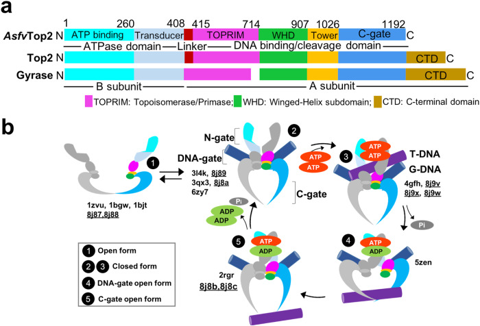
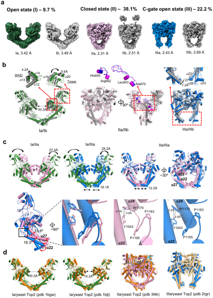
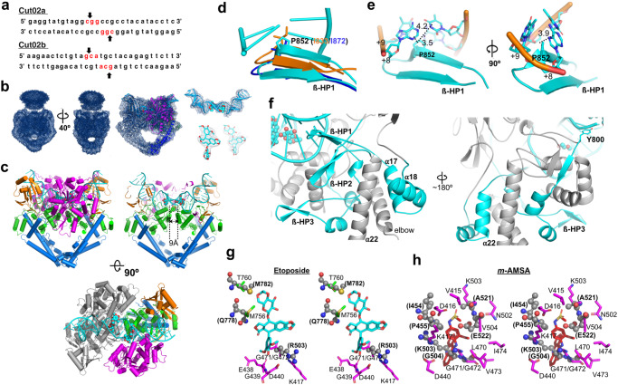
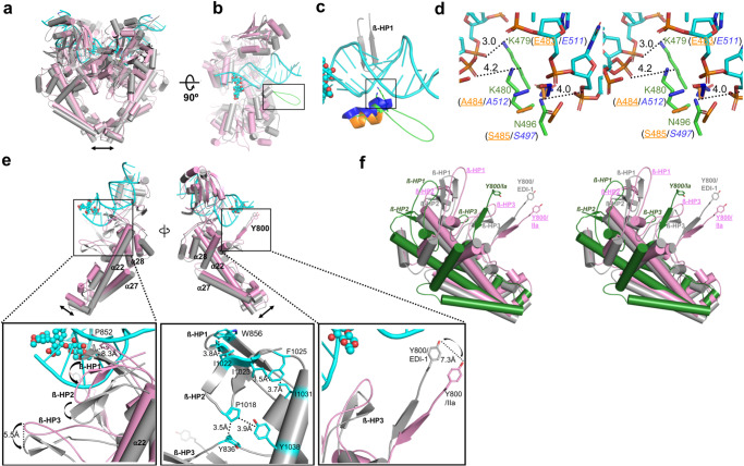
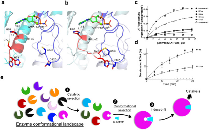
The theories for substrate recognition in enzyme catalysis have evolved from lock-key to induced fit, then conformational selection, and conformational selection followed by induced fit. However, the prevalence and consensus of these theories require further examination. Here we use cryogenic electron microscopy and African swine fever virus type 2 topoisomerase (AsfvTop2) to demonstrate substrate binding theories in a joint and ordered manner: catalytic selection by the enzyme, conformational selection by the substrates, then induced fit. The apo-AsfvTop2 pre-exists in six conformers that comply with the two-gate mechanism directing DNA passage and release in the Top2 catalytic cycle. The structures of AsfvTop2-DNA-inhibitor complexes show that substantial induced-fit changes occur locally from the closed apo-conformer that however is too far-fetched for the open apo-conformer. Furthermore, the ATPase domain of AsfvTop2 in the MgAMP-PNP-bound crystal structures coexist in reduced and oxidized forms involving a disulfide bond, which can regulate the AsfvTop2 function.


Conflict of interest statement

The authors declare no competing interests.

Figures

Fig. 1
Fig. 1. Top2 domain composition and the main conformational states in its catalytic cycle.
a Schematic illustration for the subdomains of AsfvTop2, Top2 and gyrase. b Cartoon model illustration of the proposed Top2 conformational states (numbered in black circles),,,–,,, with corresponding subdomains colored as indicated in the scheme above. The cycle starts with the apo-Top2 existing in an open dimeric form (state 1), which closes upon Gate (G) DNA binding to the DNA cleavage core (state 2). Next, the N-terminal ATPase (N-gate) is closed in the presence of ATP to capture the Transport (T) DNA segment (state 3). Then a double-strand G-DNA break occurs transiently to enable the T-DNA passage through the open DNA-gate (state 4), with one ATP molecule concurrently hydrolyzed. Next, re-ligation of the cleaved G-DNA segment triggers opening of the C-gate, leading to the release of T-DNA (state 5). Finally, the enzyme returns to the state 2 by closing the C-gate and releasing ADP. These conformational states or their variants are often referred to as the open form (state 1) or closed form (states 2–3) based on the status of the DNA-gate, while states 4 and 5 are respectively described as DNA-gate open and C-gate open forms here. The PDB codes for the previously reported structures and those from this study (Table 1) are listed, with the latter underlined.
Fig. 2
Fig. 2. Structures of six conformers from apo-AsfvTop2.
a Cryo-EM density maps for the six conformers of the full-length AsfvTop2. The selected particle percentages of conformer I, II and III are labeled. The densities for the subdomains were clearly defined at a resolution of 2.31–3.49 Å, except for the ATPase domain. Sparse densities for ATPase of conformer IIb can be seen, which indicates that these domains were not cleaved in the process of obtaining structures, but were flexible instead. The TOPRIM subdomain is not modeled in the conformer I. b High resolution cryo-EM reconstructions of the apo-AsfvTop2 cleavage core domain for the three states (I-III), with the two sub-conformers of each state superposed with each other. The distance between the Cα atoms of the Y744/Y’744 from the two α-helices 13 on the WHD subdomains of the two subunits is shown for Ia (Ia: 30.3 Å; Ib: 27.6 Å). The detailed differences between sub-conformers are shown in insets. Annotation of the helices is based on the structure-based alignment (Supplementary Figs. 5, 6). c Upper panel: Superposed structures showing major conformational changes between conformers Ia and IIa (left); Ia and IIIa (center), and IIa and IIIa (right). The TOPRIM subdomain is omitted for clarity. Lower panel: The hinge connecting helix α27 and α28 contains two non-conserved proline residues. The residues and contacts that contribute to the structural differences are labeled. d Superposition of Ia, Ib, IIa, and IIIa with the comparable crystal structures from the yeast Top2 cleavage core: pdb 1bgw (apo, TOPRIM omitted), 1bjt (apo, TOPRIM omitted), 3l4k (crossed-linked with dsDNA, closed state), and 2rgr (complexed with dsDNA, closed state with C-gate open), respectively.
Fig. 3
Fig. 3. Cryo-EM structure of AsfvTop2-DNA-drug complex showing unique DNA interactions and drug binding modes.
a The DNA sequences of the two G-DNA segments (Cut02a and Cut02b). The cleavage sites are indicated with arrows. b Left: The representative global cryo-EM density of the full-length complexes, AsfvTop2 Cut02a/inhibitor (EDI-1, resolution 2.70 Å, contoured at 0.034 σ). Center: The global map fitted with the modeled cartoon structure, contoured at 0.198 σ. Right: Maps of bound DNA and inhibitor molecules. c Left: The EDI-1 complex is shown in cartoon cylinders and colored based on the color scheme in Fig. 1a. Center/right: The structure is slabbed through to see the two α-helices 13 clearly with their distance labeled. The DNA and inhibitor are colored in cyan with cartoon and sphere presentations, respectively. d Enlarged view of the overlaid ß-HP1 region (colored in cyan, residues 846–861). The conserved isoleucine in yeast (orange)/human (blue) Top2s, and AsfvTop2 P852 (shown in stick) emanate towards DNA to different extents. e The intercalation of P852 and its distances (Å) to adjacent DNA bases are labeled. f Left: the spatial location of the ß-HP1, adjacent to ß-HP2 (residues 822-834) and ß-HP3 (residues 1012-1025), and the elbow region. Right: ß-HP1 is linked to the catalytic Y800 through ß-HP3. The structural moieties mentioned are highlighted in cyan. g, h Structural superimposition between AsfvTop2 and human Top2 showing the differences in drug binding in stereo views. The corresponding human Top2 residues (pdb 3qx3, etoposide bound, pdb 4g0u, m-AMSA bound) are shown in spheres and labeled in bold letters in parentheses.
Fig. 4
Fig. 4. Global and local conformational changes induced by DNA binding.
a The subdomain reorganization of AsfvTop2 is illustrated by aligning EDI-1 (gray) with apo-IIa (pink). b Half model of a, with a 90° rotation around the x-axis. The black rectangle highlights the extended loop that appears upon DNA binding. c The extended loop (residues 481-493, colored in green) shown inside the black square in panel b. The corresponding region in yeast/human Top2, is colored in orange/blue, respectively. d The close-up stereo view of the specific interactions between AsfvTop2 and DNA in the square box in (c). The interactions are governed by the K479 and K480 of AsfvTop2, which are replaced with Glu and Ala (residue numbers in parentheses), respectively, in yeast/human Top2. e Upper panels: A global view of the local movements upon DNA binding with focus on the ß-HP1 region (left) and the catalytic Y800 (right). Lower panels: The enlarged view of the squared regions in the upper panel. Left: The three ß-HP loops from apo-IIa concurrently shift downward in the EDI-1 complex. Middle: The hydrophobic network that coordinates the concurrent movements of the three ß-HPs and α22 in the EDI-1 complex. Right: Shifting of the catalytic Y800 toward DNA in the complex. f Stereo view for the spatial locations of the three ß-HPs and Y800 in apo-Ia (colored in green with italic label), apo-IIa (colored in pink with underlined label), and the EDI-1 complex (gray).
Fig. 5
Fig. 5. A potential redox regulatory mechanism unique to AsfvTop2.
a, b The close-up view for the relative positions of MgAMP-PNP and the essential residues (C72, C138, H68, H73, and D137, all in stick presentation) involved in the potential regulatory mechanism. The reduced (a) and oxidized (b) forms are shown in cyan and pale cyan cartoon presentations, respectively. The disulfide bond between C72 and C138 is shown in brown stick. D137 and C138 are located at a long-extended surface loop (colored in blue) that harbors the active site residues interacting with the triphosphate group of the AMP-PNP. The segment E69-H73 that moves to form the disulfide bond is shown in red. c Analysis of ATP hydrolysis for the AsfvTop2 ATPase domain in the presence of 1 mM ATP and varying concentrations of the enzyme. The y-axis shows the specific activity from each measurement. Raw data are provided in Supplementary Data 5. d C72A mutation of the full-length AsfvTop2 resulted in significant reduction of DNA decatenation activity in comparison to the wild type over different time points. Detailed conditions are described in Supplementary Fig. 13c, d. Raw data are provided in Supplementary Data 6. Each data point in panels c, d includes the mean ± SE value from three independent reactions (n = 3) from the same batch of sample. Similar assay methods for ATPase and decatenation activities have been used previously for other Top2 proteins,. e A graphical summary illustrating that the enzyme-substrate recognition is mediated by the joint application of the substrate binding theories in order: (1) catalytic selection, (2) conformational selection, and (3) induced fit. The catalytic selection should be replaced by functional selection when referring to other functions instead of catalysis.

Similar articles

Cited by

References

    1. Fischer E. Einfluss der configuration auf die wirkung der enzyme. Ber. Dtsch. Chem. Ges. 1894;27:2985–1993. doi: 10.1002/cber.18940270364. - DOI
    1. Koshland DE. Application of a theory of enzyme specificity to protein synthesis. Proc. Natl Acad. Sci. USA. 1958;44:98–104. doi: 10.1073/pnas.44.2.98. - DOI - PMC - PubMed
    1. Monod J, Wyman J, Changeux JP. On the nature of allosteric transitions: a plausible model. J. Mol. Biol. 1965;12:88–118. doi: 10.1016/S0022-2836(65)80285-6. - DOI - PubMed
    1. Ma B, Nussinov R. Enzyme dynamics point to stepwise conformational selection in catalysis. Curr. Opin. Chem. Biol. 2010;14:652–659. doi: 10.1016/j.cbpa.2010.08.012. - DOI - PMC - PubMed
    1. Weikl TR, Paul F. Conformational selection in protein binding and function. Protein Sci. 2014;23:1508–1518. doi: 10.1002/pro.2539. - DOI - PMC - PubMed