

Brain structural and functional outcomes in the offspring of women experiencing psychological distress during pregnancy
- PMID: 38418579
- PMCID: PMC11408260
- DOI: 10.1038/s41380-024-02449-0
Brain structural and functional outcomes in the offspring of women experiencing psychological distress during pregnancy
Abstract
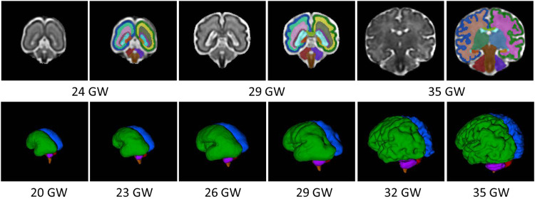
In-utero exposure to maternal psychological distress is increasingly linked with disrupted fetal and neonatal brain development and long-term neurobehavioral dysfunction in children and adults. Elevated maternal psychological distress is associated with changes in fetal brain structure and function, including reduced hippocampal and cerebellar volumes, increased cerebral cortical gyrification and sulcal depth, decreased brain metabolites (e.g., choline and creatine levels), and disrupted functional connectivity. After birth, reduced cerebral and cerebellar gray matter volumes, increased cerebral cortical gyrification, altered amygdala and hippocampal volumes, and disturbed brain microstructure and functional connectivity have been reported in the offspring months or even years after exposure to maternal distress during pregnancy. Additionally, adverse child neurodevelopment outcomes such as cognitive, language, learning, memory, social-emotional problems, and neuropsychiatric dysfunction are being increasingly reported after prenatal exposure to maternal distress. The mechanisms by which prenatal maternal psychological distress influences early brain development include but are not limited to impaired placental function, disrupted fetal epigenetic regulation, altered microbiome and inflammation, dysregulated hypothalamic pituitary adrenal axis, altered distribution of the fetal cardiac output to the brain, and disrupted maternal sleep and appetite. This review will appraise the available literature on the brain structural and functional outcomes and neurodevelopmental outcomes in the offspring of pregnant women experiencing elevated psychological distress. In addition, it will also provide an overview of the mechanistic underpinnings of brain development changes in stress response and discuss current treatments for elevated maternal psychological distress, including pharmacotherapy (e.g., selective serotonin reuptake inhibitors) and non-pharmacotherapy (e.g., cognitive-behavior therapy). Finally, it will end with a consideration of future directions in the field.
© 2024. The Author(s).
Conflict of interest statement
All authors declare no competing interests.
Figures

References
-
- Gavin NI, Gaynes BN, Lohr KN, Meltzer-Brody S, Gartlehner G, Swinson T. Perinatal depression: a systematic review of prevalence and incidence. Obstet Gynecol. 2005;106:1071–83. - PubMed
Publication types
MeSH terms
LinkOut - more resources
Full Text Sources
Medical
