. 2024 Aug;46(4):3831-3844.
doi: 10.1007/s11357-024-01091-6. Epub 2024 Feb 28.

Functional deterioration of vascular mitochondrial and glycolytic capacity in the aortic rings of aged mice

Affiliations

Functional deterioration of vascular mitochondrial and glycolytic capacity in the aortic rings of aged mice

Agnieszka Karaś et al. Geroscience. 2024 Aug.

Abstract

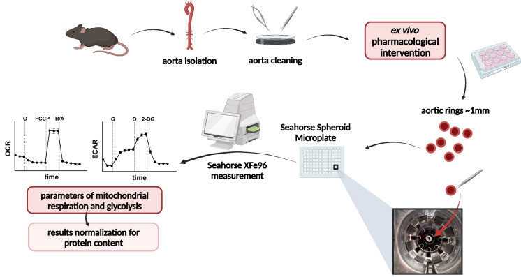
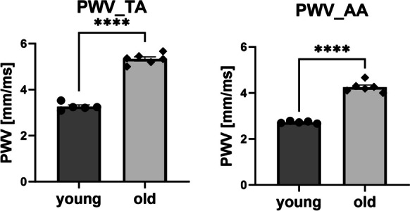
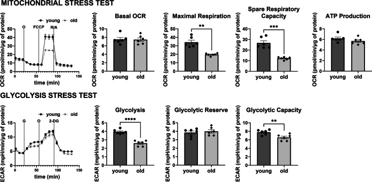
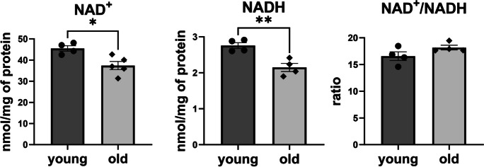
Vascular ageing is associated with increased arterial stiffness and cardiovascular mortality that might be linked to altered vascular energy metabolism. The aim of this study was to establish a Seahorse XFe96 Analyzer-based methodology for the reliable, functional assessment of mitochondrial respiration and glycolysis in single murine aortic rings and to validate this functional assay by characterising alterations in vascular energy metabolism in aged mice. Healthy young and old C57BL/6 mice were used for the analyses. An optimised setup consisting of the Seahorse XFe96 Analyzer and Seahorse Spheroid Microplates was applied for the mitochondrial stress test and the glycolysis stress test on the isolated murine aortic rings, supplemented with analysis of NAD content in the aorta. To confirm the age-dependent stiffness of the vasculature, pulse wave velocity was measured in vivo. In addition, the activity of vascular nitric oxide synthase and vascular wall morphology were analysed ex vivo. The vascular ageing phenotype in old mice was confirmed by increased aortic stiffness, vascular wall remodelling, and nitric oxide synthase activity impairment. The rings of the aorta taken from old mice showed changes in vascular energy metabolism, including impaired spare respiratory capacity, maximal respiration, glycolysis, and glycolytic capacity, as well as a fall in the NAD pool. In conclusion, optimised Seahorse XFe96-based analysis to study energy metabolism in single aortic rings of murine aorta revealed a robust impairment of functional vascular respiratory and glycolytic capacity in old mice linked to NAD deficiency that coincided with age-related aortic wall remodelling and stiffness.

Keywords: Ageing; Aorta; Glycolysis; Mitochondrial function; NAD; Seahorse XF technique; Vascular stiffness.


Conflict of interest statement

The authors declare no competing interests.

Figures

Fig. 1
Fig. 1
Graphical presentation of the experimental protocol of the measurement of vascular bioenergetics in murine isolated aortic rings using Seahorse XFe96 Analyzer (created with BioRender.com). O – oligomycin, FCCP – carbonyl cyanide 4-[trifluoromethoxy]phenylhydrazone, G – glucose, 2-DG – 2-deoxyglucose
Fig. 2
Fig. 2
Increased vascular wall stiffness measured in vivo in the aorta of old C57BL/6 mice. Aortic stiffness was assessed as pulse wave velocity (PWV) index in old (24-month-old) as compared to young (3-month-old) C57BL/6 mice in the thoracic (TA) and abdominal (AA) aorta. Data represent the means ± SEM (n = 5–6), analysed with t-test, ****P ≤ 0.001
Fig. 3
Fig. 3
Aortic wall remodelling in old C57BL/6 mice using histological analysis. Representative images of haematoxylin and eosin (HE) staining (n = 6) and collagen fibre staining using picrosirius red (PSR) (n = 6) in the aorta of old (25-month-old) (B) as compared with young (3-month-old) C57BL/6 mice (A) in 100 × magnification (top panel) and 400 × magnification (bottom panel). C Aortic media thickness measured on PSR staining images, as demonstrated with arrows in the figures A and B. Data represent means ± SEM (n = 5–6), analysed with t-test, ****P ≤ 0.0001. D Relative collagen content in media assessed as a percentage of red pixels on PSR-stained images. Data represent means ± SEM (n = 6), analysed with t-test, **P ≤ 0.01
Fig. 4
Fig. 4
Impaired NOS activity in the aorta of old C57BL/6 mice assessed by the metabolism of stable isotope labelled L-arginine. Aorta isolated from adult (8-month-old) and old (26-month-old) C57BL/6 mice was incubated for 24 h with 13C6, 15N4 L-arginine-HCl (arginine + 10) and stimulated with calcium ionophore for the last 90 min. The levels of labelled arginine and its metabolites, 13C6, 15N3 L-citrulline (citrulline + 9) and 13C5, 15N2 L-ornithine (ornithine + 7) were measured using LC/MS method and expressed as relative ratios. Data represent means ± SEM (n = 8–9), analysed with t-test/Mann–Whitney, *P ≤ 0.05
Fig. 5
Fig. 5
Alterations in functional vascular energy metabolism profile in aorta from old C57BL/6 mice as compared with young C57BL/6 mice measured in single aortic rings using the Seahorse XF Analyzer, with mitochondrial stress test (MST) and the glycolysis stress test (GST). Basal bioenergetics were assessed in aortic rings isolated from old (25-month-old) C57BL/6 male mice compared with young (3-month-old) mice using the Seahorse XFe96 Extracellular Analyzer and mitochondrial stress test protocol and glycolytic stress test. Data represent the means ± SEM (n = 6), analysed with t-test, *P ≤ 0.05, **P ≤ 0.01, ***P ≤ 0.001, ****P ≤ 0.001
Fig. 6
Fig. 6
Fall in the content of nicotinamide adenine dinucleotide (NAD) in the aorta of old C57BL/6 mice as compared with young C57BL/6 mice. Nicotinamide adenine dinucleotide concentrations in both oxidised (NAD.+) and reduced form (NADH) were assessed using HPLC method in young (4-month-old) and old (25-month-old) C57BL/6 mice in the thoracic aorta. Data represent the means ± SEM (n = 4), analysed with t-test, *P ≤ 0.05, **P ≤ 0.01

References

    1. Ungvari Z, Tarantini S, Donato AJ, Galvan V, Csiszar A. Mechanisms of vascular aging. Circ Res. 2018;123:849–867. doi: 10.1161/CIRCRESAHA.118.311378. - DOI - PMC - PubMed
    1. Lakatta EG, Levy D. Arterial and cardiac aging: major shareholders in cardiovascular disease enterprises: part I: aging arteries: a “set up” for vascular disease. Circulation. 2003;107:139–146. doi: 10.1161/01.CIR.0000048892.83521.58. - DOI - PubMed
    1. Campisi J, Kapahi P, Lithgow GJ, Melov S, Newman JC, Verdin E. From discoveries in ageing research to therapeutics for healthy ageing. Nature. 2019;571:183–192. doi: 10.1038/s41586-019-1365-2. - DOI - PMC - PubMed
    1. Tyrrell DJ, Blin MG, Song J, Wood SC, Goldstein DR. Aging impairs mitochondrial function and mitophagy and elevates interleukin 6 within the cerebral vasculature. J Am Heart Assoc 2020;9. 10.1161/JAHA.120.017820. - PMC - PubMed
    1. Fleenor BS, Seals DR, Zigler ML, Sindler AL. Superoxide-lowering therapy with TEMPOL reverses arterial dysfunction with aging in mice. Aging Cell. 2012;11:269–276. doi: 10.1111/j.1474-9726.2011.00783.x. - DOI - PMC - PubMed

Publication types

LinkOut - more resources