. 2024 Mar;627(8002):157-164.
doi: 10.1038/s41586-024-07108-6. Epub 2024 Feb 28.

Neuronal dynamics direct cerebrospinal fluid perfusion and brain clearance

Affiliations

Neuronal dynamics direct cerebrospinal fluid perfusion and brain clearance

Li-Feng Jiang-Xie et al. Nature. 2024 Mar.

Abstract

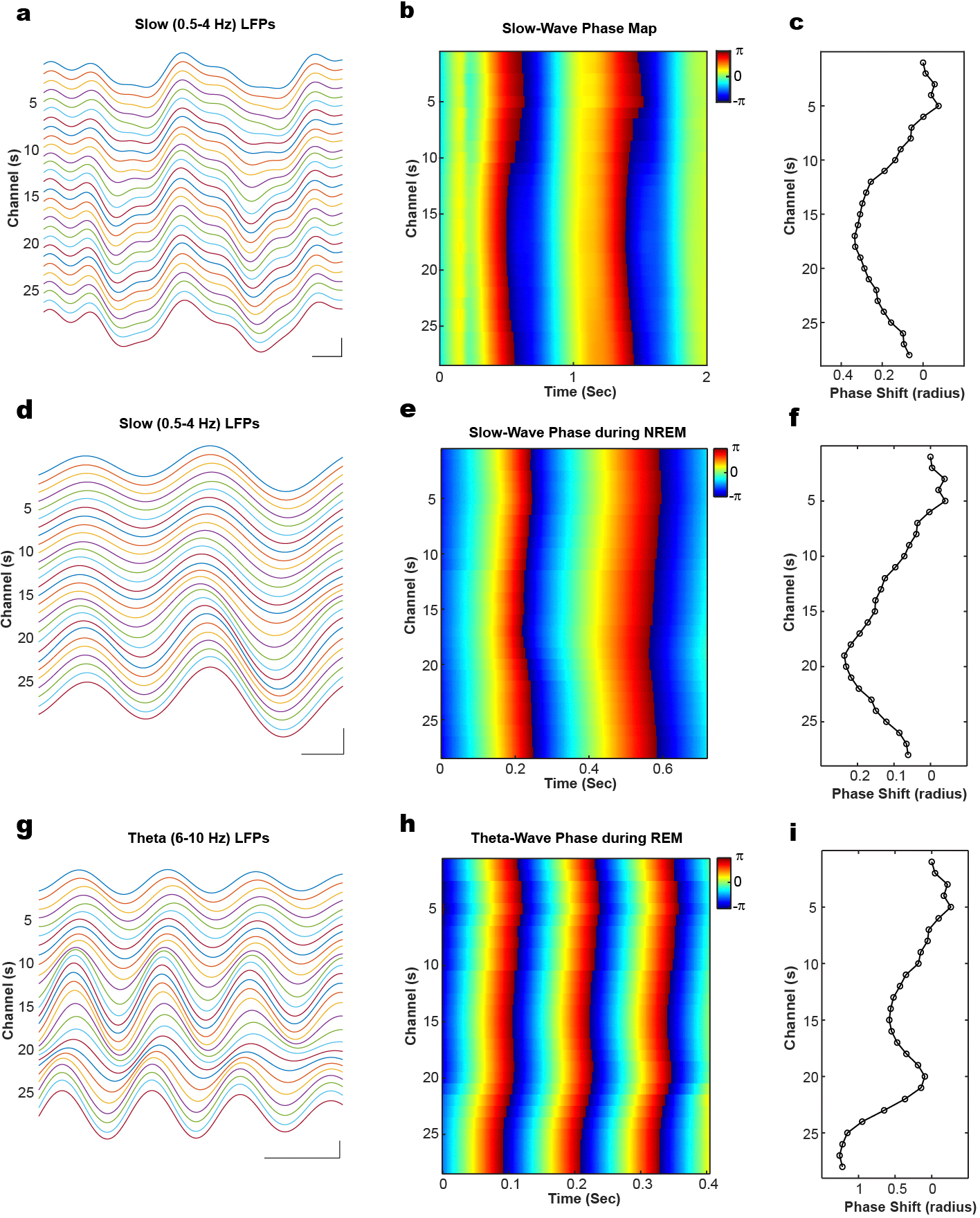
The accumulation of metabolic waste is a leading cause of numerous neurological disorders, yet we still have only limited knowledge of how the brain performs self-cleansing. Here we demonstrate that neural networks synchronize individual action potentials to create large-amplitude, rhythmic and self-perpetuating ionic waves in the interstitial fluid of the brain. These waves are a plausible mechanism to explain the correlated potentiation of the glymphatic flow1,2 through the brain parenchyma. Chemogenetic flattening of these high-energy ionic waves largely impeded cerebrospinal fluid infiltration into and clearance of molecules from the brain parenchyma. Notably, synthesized waves generated through transcranial optogenetic stimulation substantially potentiated cerebrospinal fluid-to-interstitial fluid perfusion. Our study demonstrates that neurons serve as master organizers for brain clearance. This fundamental principle introduces a new theoretical framework for the functioning of macroscopic brain waves.


Conflict of interest statement

Competing interests J.K. is a co-founder of Rho Bio. The other authors declare no competing interests.

Figures

Extended Data Fig. 1 |
Extended Data Fig. 1 |. Characterization of EEGs and ionic waves in ISF of hippocampus during wake and ketamine anesthesia with chemogenetic manipulation.
a, Schematic of electrode location in PSAM-expressed hippocampus. bi, Power spectra of frontal EEG (fEEG) and parietal EEG (pEEG) during wake, ketamine anesthesia (Ket) and Ket + uPSEM792. n = 5 mice for wake; n = 4 mice for Ket and Ket + uPSEM792. Shaded areas denote 95 percent confidence intervals for the mean. bii, Power spectra of ISF waves in hippocampus during wake, ketamine anesthesia with or without chemogenetic inhibition. n = 136 channes from 5 mice. Shaded areas denote 95 percent confidence intervals for the mean. c-e, Cross-frequency coupling analysis in hippocampal ISF during ketamine anesthesia, scale bar: 300 ms and 200 μV. Raw and filter traces (c), phase and power extraction (d), and polar plot across all 28-recording channels (e).
Extended Data Fig. 2 |
Extended Data Fig. 2 |. Analysis of neuronal spikes in hippocampus during wake and ketamine anesthesia with chemogenetic perturbation.
a, Examples of spike waveforms. b, Spike rate for isolated units during wakefulness and ketamine (n = 34 units from 5 mice). Left, statistical summary of unit spike rates; right, normalized activity heatmap. Two-sided Wilcoxon signed-rank test. p < 0.0001. c, Instant amplitude of field potential coupled with neuronal spikes (n = 34 units from 5 mice). Two-sided Wilcoxon signed-rank test. p < 0.0001. d, Spike rate for isolated units during wakefulness and ketamine with chemogenetic inhibition (n = 35 units from 5 mice). Left, statistical summary of unit spike rates; right, normalized activity heatmap. Two-sided Wilcoxon signed-rank test. p < 0.0001. *** p < 0.001.
Extended Data Fig. 3 |
Extended Data Fig. 3 |. Molecular infiltration analysis on additional brain regions.
a, Representative images from PSAM-expressing animals. Left column displays composite images (GFP, Dex, and DAPI) from anterior to posterior (top to bottom); while the right column shows corresponding tracer-only (Dex) images. Statistical summary for tracer infiltration in GFP group. (b) and PSAM group (c) across multiple brain regions: Ant DC, anterior dorsal cortex; Ant VC, anterior ventral cortex; Post DC, posterior dorsal cortex; Post VC, posterior ventral cortex; Hypo, hypothalamus. n = 7 mice for both GFP and PSAM groups. Scale bar: 500 μm. Two-sided Paired-t test. n.s., not significant.
Extended Data Fig. 4 |
Extended Data Fig. 4 |. Acute neuronal inhibition does not affect GFAP expression and blood-brain barrier integrity.
a, GFAP staining after uPSEM792 treatment (3 mg/kg, i.p.) in GFP group (left panel, n = 4) and PSAM group (right panel, n = 4). GFAP, glial fibrillary acidic protein. Scale bar: 500 μm. Paired-t test. n.s., not significant. b, Blood-brain barrier leakage assay after uPSEM792 treatment (3 mg/kg, i.p.) in GFP group (left panel, n = 4) and PSAM group (right panel, n = 4). Scale bar: 500 μm. Two-sided paired-t test. n.s., not significant.
Extended Data Fig. 5 |
Extended Data Fig. 5 |. Characterization of EEGs and ISF waves in hippocampus during natural sleep-wake cycle with chemogenetic inhibition.
a, Representative traces of EEG, EMG, and ionic waves in the hippocampus during wake, NREM, and REM with chemogenetic inhibition. Top right scale bar: 200 ms and 200 μV for EEGs; bottom right scale bar: 200 ms and 200 μV for LFPs. b, Power spectra of cortical EEGs (n = 4 animals) before and after chemogenetic inhibition. c, Power spectra of hippocampal ISF before (n = 111 channels from 4 animals) and after (n = 109 channels from 4 animals) chemogenetic inhibition. Shaded areas denote 95 percent confidence intervals for the mean.
Extended Data Fig. 6 |
Extended Data Fig. 6 |. Analysis of neuronal spikes in hippocampus during natural sleep-wake cycle with chemogenetic perturbation, the asymmetry of CSF perfusion between sleep and wake.
a, Examples of spike waveforms. b-c, Spike rate for isolated units (n = 37 units from 4 mice) underlying wake, NREM, and REM with chemogenetic inhibition. b, Statistical summary of unit spike rates; c, Normalized activity heatmap. d, Instant amplitude coupled with neuronal spikes (n = 37 units from 4 mice). Two-sided Wilcoxon signed-rank test with Bonferroni correction. p = 0.0001 (Wake vs NREM) and p < 0.0001 (Wake vs REM) e, Asymmetry of CSF perfusion between wake (n = 10 animals) and sleep (n = 9 animals) conditions measured as percentage of contralateral (Contra) side. Two-sided Student t-test. p = 0.0045. ** p < 0.01, *** p < 0.001.
Extended Data Fig. 7 |
Extended Data Fig. 7 |. EEG/EMG characterization after acute intracisterna magna (ICM) injection.
a, Representative spectrograms from fEEG (top row), pEEG (second row), EMG (third row), hypnogram (fourth row) after acute ICM injection. b, Representative recording traces in three different brain states, wake, NREM, and REM. Blue trace, fEEG; red trace, pEEG, black trace, EMG. Scale bar: 500 ms and 200 μV. c, Power spectrum analysis across wake, NREM, and REM in the frontal EEG (top) and parietal (bottom) EEG channels. d, Percentage of time spent in Wake, NREM, and REM sleep., n = 3 animals. fEEG, frontal EEG; pEEG, parietal EEG.
Extended Data Fig. 8 |
Extended Data Fig. 8 |. MRI, the time window for molecular clearance, and the efficacy and duration of chemogenetic inhibition.
a-b, Paravascular flow is largely preserved during local neuronal inhibition in hippocampus. Left panel, GFP group; right panel, PSAM group. GadoSpinP, large molecular tracer (~200 kDa) used to visualize para-vascular flow. ICM, intracisterna magna injection. n = 3 mice for GFP group; n = 4 mice for PSAM group. Data presented as mean ± s.e.m. c, 3–7 h after ICM (3 kD Dextran-TexasRed, yellow) injection mainly captures molecular clearance phase. n = 6 mice for 3-hour group; n = 8 mice for 7-hour group. Two-sided Mann-Whitney test, p = 0.0426. Scar bar = 500 μm. d, Chemogenetic inhibition lasts for 5 h after uPSEM792 injection across three brain states, wake, NREM, REM. n = 109 channels from 4 animals. Shaded areas denote 95 percent confidence intervals for the mean. *p < 0.05.
Extended Data Fig. 9 |
Extended Data Fig. 9 |. Validation of optogenetic toolkits.
a, Transcranial activation of neurons revealed by Fos staining. Left, representative images; right, statistical summary (n = 4 animals). Contra, contralateral side of the hippocampus. Scale bar: 500 μm. Two-sided paired-t test. p = 0.0031. b, Representative field potential traces with photo-stimulations in ChRmine-expressing animals. Slow stimulation: 1 Hz, 50 ms per TTL pulse; theta stimulation: 8 Hz, 6.25 ms per TTL pulse. Scale bar: 200 ms and 300 μV. c, Quantification of slow wave (0.5–4 Hz) power and theta wave (6–10 Hz) power from optrode recording experiment (n = 54 channels from 2 animals). d, Illustration of the principle that neurons firing together shower together. ** p < 0.01.
Extended Data Fig. 10 |
Extended Data Fig. 10 |. Wave phase progression analysis across electrophysiological recording channels in hippocampus during different brain states.
a-c, Representative slow (0.5–4 Hz) filtered traces (a) from hippocampal field potential recordings during ketamine anesthesia, corresponding phases (b) extracted with Hilbert method, and averaged phase shift (c). Scale bar: 200 ms and 200 μV. d-f, Representative slow (0.5–4 Hz) filtered traces (d) from hippocampal field potential recordings during NREM sleep, corresponding phases (e) extracted with Hilbert method, and averaged phase shift (f). Scale bar: 100 ms and 200 μV. g-i, Representative theta (6–10 Hz) filtered traces (g) from hippocampal field potential recordings during REM sleep, corresponding phases (h) extracted with Hilbert method, and averaged phase shift (i). Scale bar: 100 ms and 200 μV.
Fig. 1 |
Fig. 1 |. Large-amplitude and rhythmic ionic dynamics in ISF generated by neuronal synchronization during ketamine anaesthesia are required for brain CSF perfusion.
a, Top, illustration of in vivo multiplexed electrophysiological recording. Bottom, representative recording of ionic flow in ISF of the hippocampus. b, Representative traces of EEG, EMG and ionic waves in the hippocampus during wake (left), ketamine anaesthesia (middle) and ketamine anaesthesia with chemogenetic inhibition (right) conditions. Ketamine anaesthesia was 100 mg kg−1 ketamine with 10 mg kg−1 xylazine, i.p. injection. Top right scale bar, 200 ms and 200 μV for EEG; bottom right scale bar, 200 ms and 200 μV for local field potentials (LFPs). D, dorsal; V, ventral. c, Representative spike-triggered field potential averaging during wake and ketamine anaesthesia conditions. The unit for the colour bar is μV. d, Summary of all spiking units (n = 34 units from 5 mice) and their relationship with the extracted amplitudes (Hilbert method) of field potentials. The unit for the colour bar is μV. e, Schematic of fluorescent CSF-to-ISF tracing with or without chemogenetic inhibition. Dex, dextran–Texas Red, 3 kDa. f,g, Images (left) and quantification (right) of CSF tracer infiltration in mice expressing control GFP (f; n = 7 mice) or PSAM (g; n = 7 mice). For the representative images, the left column displays composite images (GFP, Dex and DAPI) from anterior to posterior (top to bottom), whereas the right column demonstrates corresponding tracer-only (Dex) images. Contra, contralateral side of the hippocampus. White dashed lines highlight bilateral hippocampi. Scale bar, 500 μm. Two-sided paired t-test. P = 0.0003 in PSAM group comparison. a.u., arbitrary units; NS, not significant; ***P < 0.001.
Fig. 2 |
Fig. 2 |. Synchronization of neuronal pumps during sleep drives high-energy ionic waves in ISF, which enhances CSF infusion.
a, Representative traces of EEG, EMG and ionic waves in the hippocampus during wake, NREM and REM conditions. Top right scale bar, 200 ms and 200 μV for EEG; bottom right scale bar, 200 ms and 200 μV for LFPs. b, Representative spike-triggered field potential averaging during wake, NREM and REM conditions. The unit for the colour bar is μV. c, Summary of all spiking units (n = 37 units from 4 mice) and their relationship with the extracted amplitudes (Hilbert method) of field potentials. The unit for the colour bar is μV. d, Schematic of fluorescent CSF-to-ISF tracing with or without chemogenetic inhibition during sleep and wakefulness. e, The differentiated effect of chemogenetic inhibition during wakefulness and sleep. For each brain state, the left column displays composite images (GFP, Dex and DAPI) from anterior to posterior (top to bottom), whereas the right column demonstrates corresponding tracer-only (Dex) images. White dashed lines highlight bilateral hippocampi. Scale bar, 500 μm. f, CSF tracer intensity in whole brain slices (left) and unsilenced hippocampus (Hipp; right) under wake (n = 10 animals) and sleep conditions (n = 9 animals). Two-sided Mann-Whitney test. P = 0.0435 (whole slice) and 0.0172 (unsilenced hippocampus). g, Normalized tracer intensity between PSAM-expressing and contralateral hippocampi under wake (left, n = 10 mice) and sleep conditions (right, n = 9 mice). Two-sided paired t-test. P = 0.0031 (wake) and 0.0003 (sleep). *P < 0.05, **P < 0.01, ***P < 0.001.
Fig. 3 |
Fig. 3 |. Flattening ionic waves in ISF impedes brain perfusion in vivo.
a, Schematic of experimental procedures. Dotarem, gadolinium-based MRI contrast agent. b,c, CSF-to-ISF perfusion dynamics across time in control GFP-expressing (b) and PSAM-expressing (c) animals. Top, representative MRI images. Bottom, statistical summary. Red dashed lines highlight bilateral hippocampi. A, anterior; L, left; P, posterior; R, right. The viral vector was injected into the right side of the hippocampus. n = 8 (GFP) or 9 mice (PSAM). Data presented as the mean ± s.e.m. Two-way repeated-measures analysis of variance (ANOVA). P = 0.0066 for PSAM group comparison. **P < 0.01.
Fig. 4 |
Fig. 4 |. Chemogenetic inhibition of neural activity impairs brain clearance.
a, Illustration of the glymphatic influx and efflux of designated molecules and the prediction from our working hypothesis. b, Schematic of viral constructs, stereotaxic injection and experimental procedures. c,d, Molecular clearance in control GFP-expressing (c) and PSAM-expressing (d) animals. For the representative images, the left column displays composite images (GFP, Dex and DAPI) from anterior to posterior (top to bottom) of the brain, whereas the right column demonstrates corresponding Dex-only images. White dashed lines highlight bilateral hippocampi. n = 8 (GFP) and 10 (PSAM) mice. Scale bar, 500 μm. Two-sided paired t-test. P = 0.0009 for PSAM group comparison. ***P < 0.001.
Fig. 5 |
Fig. 5 |. Synthesized brain waves generated by transcranial optogenetic potentiate brain CSF-to-ISF perfusion.
a, Schematic of viral constructs, stereotaxic injection and experimental procedures. b, Rhythmic 1 Hz optogenetic stimulation on CSF-to-ISF infiltration. Left, RFP group (n = 7 mice). Right, ChRmine group (n = 9 mice). Within each panel, the left two columns display composite images (RFP or ChRmine with DAPI) from anterior to posterior (top to bottom) in vector-expressing and contralateral sides of the hippocampus, whereas the right two columns demonstrate corresponding tracer-only (Dex) images. Statistical analysis provided on the right of each panel. c, Rhythmic 8 Hz optogenetic stimulation on CSF-to-ISF infiltration. Left, RFP group (n = 8 mice). Right, ChRmine group (n = 10 mice). Within the panel, the left two columns show composite images (RFP or ChRmine with DAPI) from ipsilateral and contralateral sides of the hippocampus, whereas the right two columns illustrate the corresponding Dex images. White dashed lines highlight bilateral hippocampi. Scale bar, 500 μm (b,c). Two-sided paired t-test. P = 0.0016 (slow wave group) and P = 0.0027 (theta wave group). **P < 0.01.

Comment in

References

    1. Iliff JJ et al. A paravascular pathway facilitates CSF flow through the brain parenchyma and the clearance of interstitial solutes, including amyloid β. Sci. Transl. Med. 4, 147ra111 (2012). - PMC - PubMed
    1. Xie L et al. Sleep drives metabolite clearance from the adult brain. Science 342, 373–377 (2013). - PMC - PubMed
    1. Magistretti PJ & Allaman I Lactate in the brain: from metabolic end-product to signalling molecule. Nat. Rev. Neurosci. 19, 235–249 (2018). - PubMed
    1. Laughlin SB, de Ruyter van Steveninck RR & Anderson JC The metabolic cost of neural information. Nat. Neurosci. 1, 36–41 (1998). - PubMed
    1. Kaplan L, Chow BW & Gu C Neuronal regulation of the blood–brain barrier and neurovascular coupling. Nat. Rev. Neurosci. 21, 416–432 (2020). - PMC - PubMed

LinkOut - more resources