. 2024 Apr;628(8006):204-211.
doi: 10.1038/s41586-024-07130-8. Epub 2024 Feb 28.

Compartmentalized ocular lymphatic system mediates eye-brain immunity

Affiliations

Compartmentalized ocular lymphatic system mediates eye-brain immunity

Xiangyun Yin et al. Nature. 2024 Apr.

Abstract

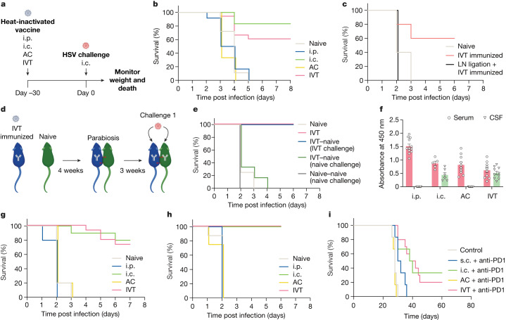
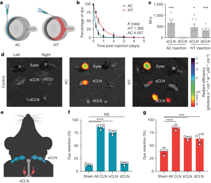
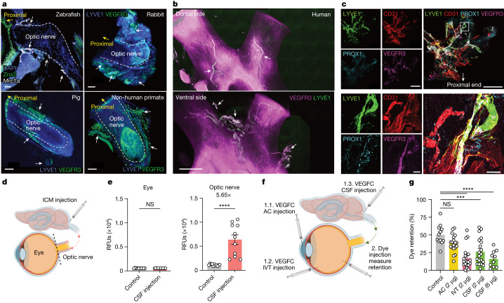
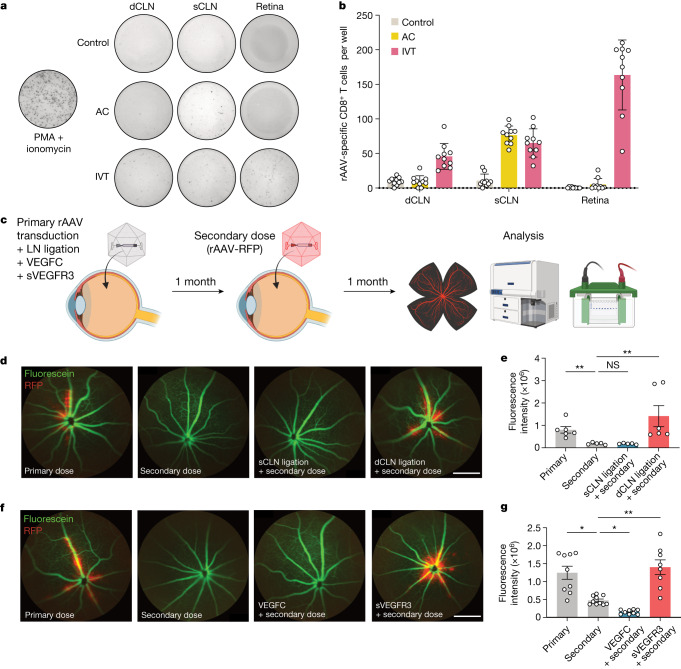
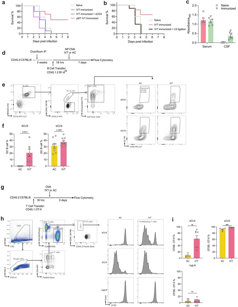
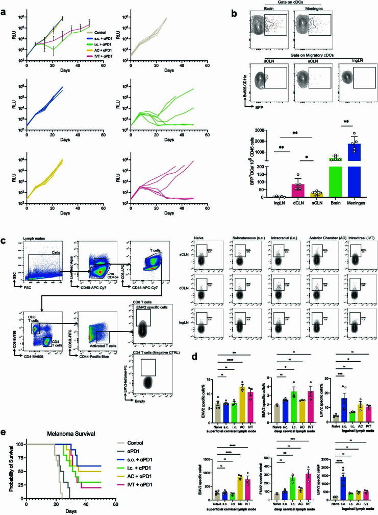
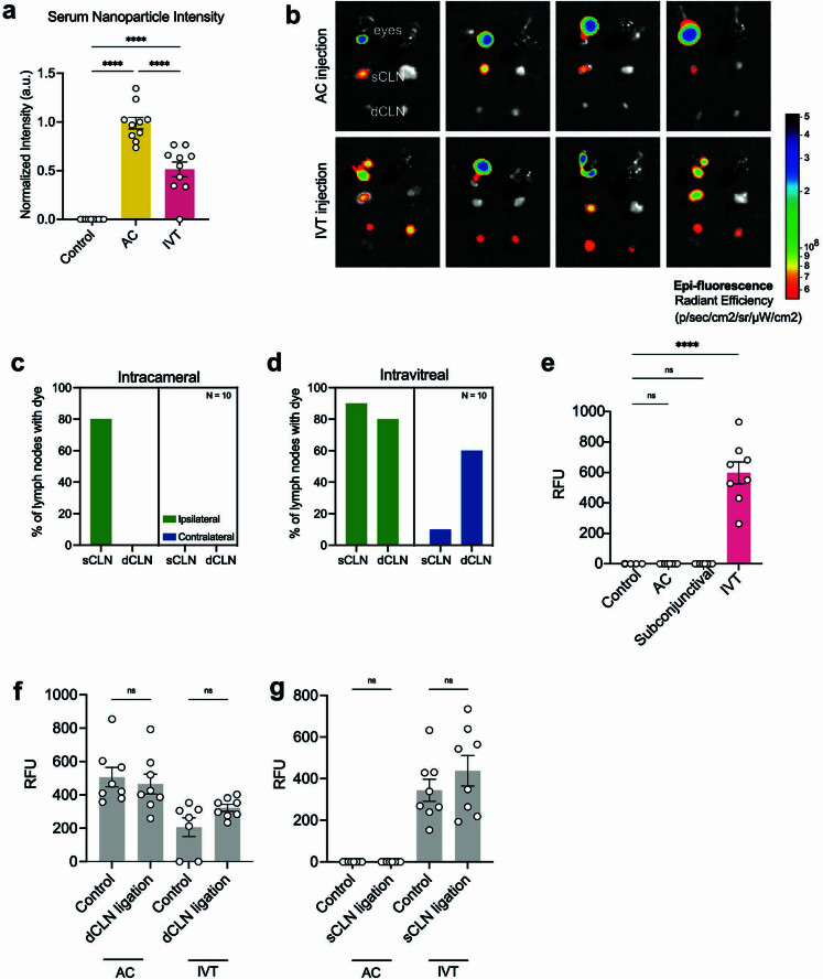
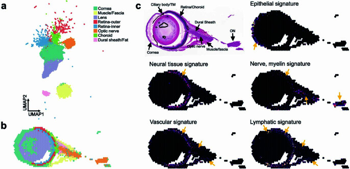
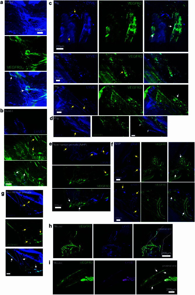
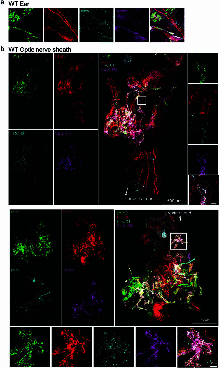
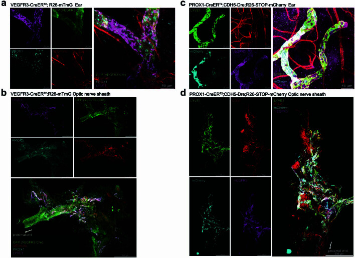
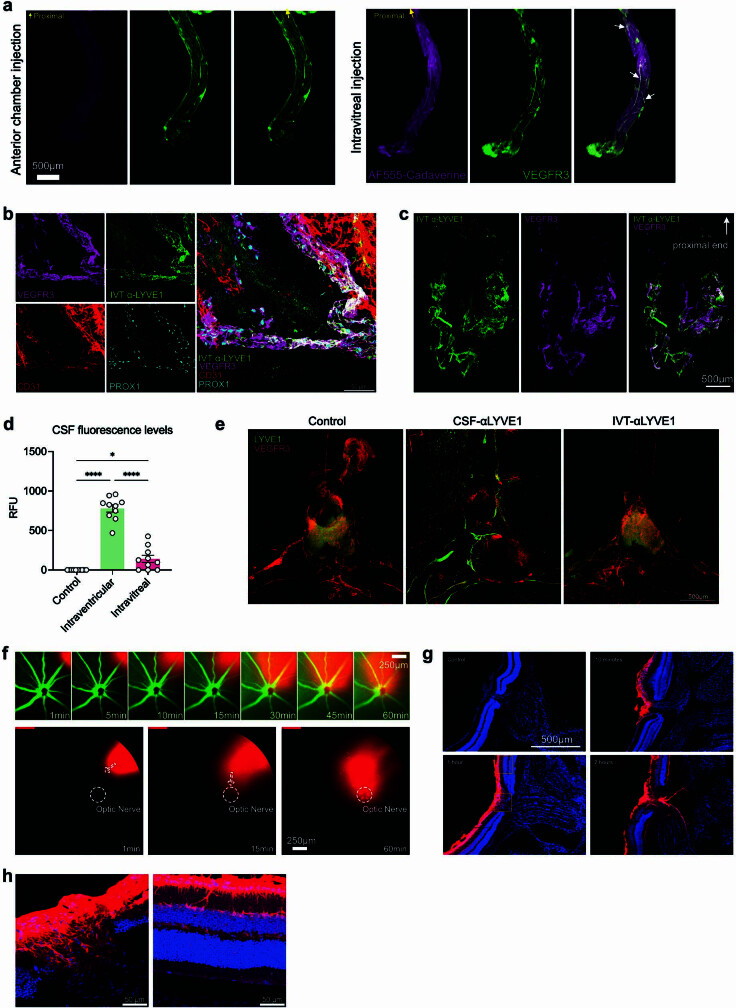
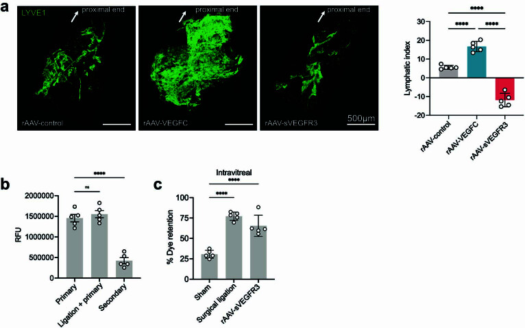
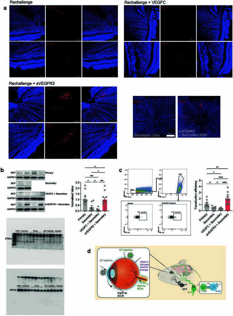
The eye, an anatomical extension of the central nervous system (CNS), exhibits many molecular and cellular parallels to the brain. Emerging research demonstrates that changes in the brain are often reflected in the eye, particularly in the retina1. Still, the possibility of an immunological nexus between the posterior eye and the rest of the CNS tissues remains unexplored. Here, studying immune responses to herpes simplex virus in the brain, we observed that intravitreal immunization protects mice against intracranial viral challenge. This protection extended to bacteria and even tumours, allowing therapeutic immune responses against glioblastoma through intravitreal immunization. We further show that the anterior and posterior compartments of the eye have distinct lymphatic drainage systems, with the latter draining to the deep cervical lymph nodes through lymphatic vasculature in the optic nerve sheath. This posterior lymphatic drainage, like that of meningeal lymphatics, could be modulated by the lymphatic stimulator VEGFC. Conversely, we show that inhibition of lymphatic signalling on the optic nerve could overcome a major limitation in gene therapy by diminishing the immune response to adeno-associated virus and ensuring continued efficacy after multiple doses. These results reveal a shared lymphatic circuit able to mount a unified immune response between the posterior eye and the brain, highlighting an understudied immunological feature of the eye and opening up the potential for new therapeutic strategies in ocular and CNS diseases.


Conflict of interest statement

E.S., A.R. and A.I. are co-founders of Rho Bio. A.I. is a member of the Board of Directors of Roche Holding Ltd. E.S., J.-L.T. and A.I. are co-inventors on patent application No. 62/929,527, “Manipulation of meningeal lymphatic vasculature for brain and CNS tumour therapy”. W.M.S. is a co-founder of B3 Therapeutics, Stradefy and Xanadu Bio. W.M.S. is a consultant to Xanadu Bio, Stradefy Biosciences, Johnson & Johnson, Celanese, Cranius and CMC Pharma.

Figures

Fig. 1
Fig. 1. Antigens in the posterior eye elicit immune responses in the brain.
a, Schematic of the schedule of procedures for the experiments described below. b, Wild-type C57BL/6J mice were immunized using heat-inactivated HSV-2 injection through i.p., i.c., AC and IVT administration. Survival was monitored after i.c. challenge with a lethal dose of HSV-2 30 days later (naive, n = 18; i.p., n = 12; i.c., n = 6; AC, n = 6; IVT, n = 18). c, dCLNs of mice were ligated using a cauterizer. Seven days later, mice were injected through the IVT route with heat-inactivated HSV-2. Their survival was monitored after i.c. challenge with a lethal dose of HSV-2 30 days later (naive, n = 5; IVT immunized, n = 5; LN ligation, n = 6). d, Schematic of the parabiosis mouse model and treatment plans. e, Mice were injected through the IVT route with heat-inactivated HSV-2. Four weeks later, the immunized mice were joined to naive mice. The immunized mice or naive mice were challenged through the i.c. route with a lethal dose of HSV-2 after 3 weeks, and their survival was monitored (naive, n = 4; IVT, n = 4; IVT–naive (IVT challenge), n = 6; IVT–naive (naive challenge), n = 2; naive–naive (naive challenge), n = 2). f, Anti-HSV-specific antibody was measured by enzyme-linked immunosorbent assay after different routes of HSV-2 immunization (i.p., n = 12; i.c., n = 6; AC, n = 10; IVT, n = 10). Data are shown as mean ± s.e.m. g, Wild-type C57BL/6J mice were injected with heat-inactivated HSV-1 through i.p., i.c., AC or IVT administration. Their survival was monitored after i.c. challenge with a lethal dose of HSV-1 30 days later (naive, n = 15; i.p., n = 6; i.c., n = 6; AC, n = 6; IVT, n = 18). h, As in a, but S.pneumoniae strain TIGR4 was used (naive, n = 8; i.p., n = 5; i.c., n = 5; AC, n = 8; IVT, n = 8). i, Mice were inoculated through the i.c. route with 50,000 GL261 luciferase-expressing (GL261–Luc) brain tumour cells, treated with irradiated GL261–Luc cells through s.c., i.c., AC or IVT administration (day 7) along with anti-PD1 (RMP1-14) antibodies (days 7, 9 and 11) and monitored for survival (naive, n = 6; s.c., n = 6; i.c., n = 6; AC, n = 6; IVT, n = 12). Data are representative of two independent experiments. The graphics in a,d were created with BioRender.com. Source Data
Fig. 2
Fig. 2. Eyes have a compartmentalized lymphatic drainage system.
a, Schematic of AC and IVT injection of dye. b, C57BL/6J mice were injected with dye through the AC or IVT route. The percentage of dye retention in the eye was analysed from 6 h to day 5 post injection (AC, n = 6; IVT, n = 6). c, Dye was injected into the eye through the AC or IVT route. sCLNs and dCLNs were collected and measured using a fluorescence plate reader 1 h after injection. RFU, relative fluorescence unit (AC, n = 8; IVT, n = 8). Data are shown as mean ± s.e.m. ***P = 0.0002, AC sCLN; *P = 0.0167, IVT sCLN; ***P = 0.0009, IVT dCLN. d, Dye was injected into the left eye through the AC or IVT route, and eyes, sCLNs and dCLNs were collected for IVIS epifluorescence imaging. Representative background-subtracted heat maps of dye in the eye, sCLNs and dCLNs 1 h after injection are shown. e, Schematic of the anatomical locations of the sCLNs and dCLNs. f,g, sCLNs, dCLNs or both CLNs were surgically ligated. Two days later, dye was injected into the eye through the AC (f) or IVT (g) route. The percentage of dye retention in the eye was measured 12 h later. Data are shown as mean ± s.e.m. in f and g (sham AC, n = 3; n = 4 in all other conditions). ****P < 0.0001; ***P = 0.0002, IVT sCLN; ***P = 0.0003, IVT dCLN; NS, not significant. P values were calculated using a one-way analysis of variance (ANOVA) with multiple comparisons testing (Dunnett). The graphics in a,e were created with BioRender.com. Source Data
Fig. 3
Fig. 3. Optic nerve sheath lymphatics drain the posterior eye.
a,b, Immunostaining of sections of optic nerve from zebrafish with lymphatics labelled for Mrc1a (white arrows; a, top left) and iDISCO immunolabelling of meningeal lymphatic vessels of rabbit (a, top right), pig (a, bottom left), non-human primate (a, bottom right) and human (b) optic nerve and chiasma, with lymphatics showing colocalization of LYVE1 and VEGFR3 (white arrows). DAPI, 4′,6-diamidino-2-phenylindole. c, Whole-mount wild-type mouse optic nerve sheaths stained for LYVE1, CD31, PROX1 and VEGFR3. The images at the bottom show a higher-magnification view of the area highlighted in the merged image at the top right. Scale bars, 50 μm (a, top left; c, bottom merged), 500 μm (a, top right; c, top merged), 1,000 μm (a, bottom left and right) and 3,000 μm (b). d, Schematic of ICM injection and the eye and optic nerve dissection for e. e, Dye was ICM injected, and fluorescence signal intensity was measured 1 h later in the eye and optic nerve (control, n = 11; CSF injection, n = 11). ****P < 0.0001. f, Schematic of injection methods for g. g, VEGFC was injected through the AC, IVT or ICM administration route. Two days later, dye was IVT injected into the eye, and the percentage of dye retention was measured 12 h after dye injection (control, n = 12; AC, n = 19; IVT, n = 20; CSF (2 μg), n = 20; CSF (6 μg), n = 10) ****P < 0.0001; ***P < 0.0002. Data are shown as mean ± s.e.m. P values were calculated using a one-way ANOVA with multiple comparisons testing (Dunnett) or two-tailed unpaired Student’s t-test. The graphics in d,f were created with BioRender.com. Source Data
Fig. 4
Fig. 4. Lymphatic inhibition enables repeat rAAV administration.
a,b, Mice were injected with rAAV-RFP through the IVT or AC route. Their dCLNs, sCLNs and retinas were collected 10 days later, and rAAV-specific immune responses were quantified using an ELISpot assay. For cg, C57BL/6J mice were IVT injected with rAAV, and, 1 month later, were rechallenged with rAAV-RFP. The efficiency of rAAV-RFP transduction was analysed by imaging 1 month later. c, Schematic of experimental plans. d, In vivo fluorescence fundus imaging to visualize vessels (green) and RFP transduction (red) in different LN ligation conditions. Scale bar, 500 μm. e, Quantification of RFP intensity from d (primary, n = 6; secondary, n = 5; sCLN ligation, n = 5; dCLN ligation, n = 6). **P = 0.0095, primary versus secondary; **P = 0.0044, secondary versus dCLN ligation. f, In vivo fluorescence fundus imaging to visualize vessels (green) and RFP transduction (red) with addition of VEGFC or sVEGFR3. Scale bar, 500 μm. g, Quantification of RFP intensity from f (primary, n = 9; secondary, n = 10; VEGFC, n = 9; sVEGFR3, n = 8). *P = 0.0167, primary versus secondary; *P = 0.0334, secondary versus VEGFC; **P = 0.0096, secondary versus sVEGFR3. P values were calculated using a one-way ANOVA with multiple comparisons testing (Dunnett). Data are shown as mean ± s.e.m in b, e and g. The graphics in c were created with BioRender.com. Source Data
Extended Data Fig. 1
Extended Data Fig. 1. Immunization through the posterior compartment of the eye can induce an immunological protection in the brain.
a, Similar to Fig. 1a, but B cell-deficient mice (μMT) were IVT injected with heat-inactivated HSV-2 and one group of WT IVT immunized mice were treated with CD4 depletion antibody before challenge. b, Similar to Fig. 1c, dCLNs were ligated after priming of the mice but before the intracranial rechallenge. Vaccination was completed first. Then, 7 days before the rechallenge, dCLNs were ligated, and mice survival was monitored after i.c. challenge with a lethal dose of HSV-2. (Naïve, n = 5; IVT immunized, n = 6; LN ligation, n = 9). c, Extending findings from Fig. 1d, the serum and CSF of both IVT immunized mice and naive mice were collected 3 weeks after parabiosis. Anti-HSV specific antibody was measured by ELISA (n = 6). d, Schematic of experimental plans. CD45.2 C57BL/6J recipient mice were primed by immunization. 2 weeks later, CD45.1.2 B1-8hi B cells labelled with CellTrace™ Violet Cell Proliferation Kit were transferred intravenously into recipient mice. 18 h later, the mice were immunized with 10 μg NP–OVA though IVT or AC injection. Igλ+ light chain positive B1-8hi cell proliferation and germinal center (GC) formation in dCLNs and sCLNs were analyzed at day 7. e, Flow cytometry gating strategy for B1-8hi GC B cells. f, Quantification of B1-8 GC B cells in dCLNs and sCLNs (n = 7–10). g, Schematic of experimental plans. CD45.1 OVA specific CD4+ T cells labelled with CFSE were transferred intravenously into CD45.2 recipient mice. 18 h later, the mice were immunized with 50 μg OVA plus 1 μg Poly(I:C) though IVT or AC injection. FTY720 was i.p. injected to inhibit the circulation of primed T cells 24 h after immunization. The OVA-specific CD4+ T cell proliferation in dCLNs and sCLNs was analyzed 72 h after immunization. h, Flow cytometry gating strategy for analyzing OVA-specific CD4+ T proliferation (n = 5–8). i, Quantification of CFSE-negative OVA-specific CD4+ T cells in dCLNs, sCLNs, and ingLNs. Data shown as mean ± s.e.m. P-values were calculated using a two-tailed unpaired Student’s t-test. The graphics in d were created with BioRender.com. Source Data
Extended Data Fig. 2
Extended Data Fig. 2. Immunization through the posterior compartment of the eye can provide therapeutic treatment of brain tumours.
a, Mice were i.c. inoculated with 50,000 GL261-Luc cells and mice were treated with irradiated GL261-Luc through the routes of s.c., i.c., AC, or IVT administration (day 7) along with anti-PD-1 antibodies (days 7, 9 and 11) and monitored for tumour growth. RLU, relative luminescence units. (s.c., n = 6; i.c., n = 6; AC, n = 6; IVT, n = 6). b, Mice were i.c. inoculated with 50,000 CT2A-BFP cells. The percentage of BFP+ DCs in the tumour, ingLNs, meninges, dCLNs and sCLNs were analyzed 14 days after tumour injection (n = 5). c,d, Similar experiment as a. The percentage of EMV-2-specific CD8T cells were analyzed in dCLN, sCLN and ingLN at day 14. c, Flow cytometry gating strategy for EMV-2-specific CD8T cells. d, Quantification of EMV-2-specific CD8+ T cells in dCLNs, sCLNs and ingLNs. (Naïve n = 5; s.c., n = 5; i.c., n = 3; AC, n = 3; IVT, n = 3) e, Mice were s.c. inoculated with 50,000 B16 cells, treated with irradiated B16 cells through the routes of s.c., i.c., AC, IVT administration (day 7) along with anti-PD-1 antibodies (days 7, 9 and 11) and monitored for survival. (n = 10 for all groups). Data shown as mean ± s.e.m. P-values were calculated using a one-way ANOVA with multiple comparisons testing (Dunnett). Source Data
Extended Data Fig. 3
Extended Data Fig. 3. Eyes have a compartmentalized lymphatic drainage system.
a, Nanoparticles were AC or IVT injected into the eye. Serum was collected, placed on a slide and imaged on a fluorescent microscope 1 h later after injection (n = 10). b, Dye was AC or IVT injected into the left eye. Eyes, sCLNs and dCLNs were collected for IVIS epifluorescence imaging. Representative background-subtracted heat maps of dye in the eye, sCLNs and dCLNs 1 h later after injection. c,d, Dye was AC (c) or IVT(d) injected into the eye and dye presence was measured using a fluorescent plate reader. The percentage of ipsilateral and contralateral LNs with dye was measured 12 h later (n = 10 mice). e, Dye was AC, IVT, or subconjunctivally injected into the left eye. dCLNs were collected and measured using a fluorescent plate reader 1 h later after injection (n = 8). f,g, dCLNs (f) or sCLNs (g) were surgically ligated. 2 days later, dye was AC or IVT injected into the eye. sCLNs (f) or dCLNs (g) were collected and measured using a fluorescent plate reader 1 h later after injection. RFU, relative fluorescence unit. (AC, n = 8; IVT, n = 8). P-values were calculated using a one-way ANOVA with multiple comparisons testing (Dunnett). Data shown as mean ± s.e.m. P-values were calculated using a one-way ANOVA with multiple comparisons testing (Dunnett) or two-tailed unpaired Student’s t-test. Source Data
Extended Data Fig. 4
Extended Data Fig. 4. 10x VISIUM Spatial sequencing of the mouse globe and optic nerve.
Mouse eyes were processed using traditional FFPE-based histology preparation. Samples were processed using the 10x VISIUM platform and analyzed using Seurat. a, UMAP of different spatial sequencing blocks with categorization into overall tissue level structure. b, Projection of tissue-type clusters onto the globe and surrounding structures. c, Image of H&E staining and respective cell type gene signature projected onto spatial sequencing data (yellow arrows denote where the gene signature appears for given signature).
Extended Data Fig. 5
Extended Data Fig. 5. Anatomical characterization of meningeal lymphatic vessels covering optic nerve.
Additional images of VEGFR3 and LYVE1 staining on optic nerves of rabbits (a,b), pigs (c,d), non-human primates (e,f,g), and mice (h,i). Yellow arrows indicate vascular structures with single staining for either LYVE1 or VEGFR3. White arrows indicate co-stained vascular structures.
Extended Data Fig. 6
Extended Data Fig. 6. Detailed analysis of optic nerve sheath lymphatics.
a, Whole-mount murine ventral ear sheet stained with LYVE1, CD31, PROX1 and VEGFR3. b, Whole-mount of optic nerve sheaths of WT mice. Top: LYVE1, CD31, PROX1 and VEGFR3 were co-stained. Bottom: LYVE1, PDPN, PROX1 and VEGFR3 were co-stained.
Extended Data Fig. 7
Extended Data Fig. 7. Detailed analysis of optic nerve sheath lymphatics with lymphatic reporter mice.
a, b, Whole-mount murine ventral ear sheet and optic nerve sheaths of VEGFR3-CreERT2;R26- mTmG mice that were stained with LYVE1 and PROX1 (a, scale: 50 μm; b, scale: 500 μm). c, d, Whole-mount murine ventral ear sheet and optic nerve sheaths of PROX1-CreERT2;CDH5-Dre;R26-STOP-mCherry mice that were stained with LYVE1, CD31 and VEGFR3 (c, scale: 50 μm; d, scale: 500 μm).
Extended Data Fig. 8
Extended Data Fig. 8. Optic nerve sheath lymphatics drain the posterior eye.
a, Whole-mount murine optic nerve VEGFR3 staining and tracer signal on optic nerve 1 h later after AC (left) or IVT (right) injection. White arrowheads point to co-localization of VEGFR3 and tracer. b, c, Anti-LYVE1 antibody was IVT injected into the vitreous humor. Optic nerve sheaths were harvested 2 h later and co-stained with CD31, PROX1 and VEGFR3. d, Dye was IVT injected into the eye or intraventricularly into the brain. CSF was collected through the cisterna magna and measured using a fluorescent plate reader 1 h later after injection. e, Anti-LYVE1 antibody was injected IVT into the eye or ICM into the CSF. Dura was collected 2 h after injection and LYVE1 antibody binding on VEGFR3 positive lymphatic vessels was analyzed by immunofluorescence. Mice without anti-LYVE1 antibody injection were used as the control. f, AF647-OVA was IVT injected into the vitreous humor. Fluorescein was i.p. injected to label blood vessels. The kinetics of AF647-OVA drainage was tracked with Phoenix MICRON® IV imaging microscope. g, AF647-OVA was IVT injected into the vitreous humor. The eyes with optic nerve were harvested, and the location of AF647-OVA was analyzed at the indicated time points. h, Zoomed in images of g. Source Data
Extended Data Fig. 9
Extended Data Fig. 9. Lymphatic inhibition enables repeated rAAV administrations.
a, Mice were IVT injected with rAAV-VEGFC, rAAV-sVEGFR3 or rAAV-control. Two months later, their optic nerve sheaths were harvested and stained with LYVE1. b, Similar to Fig. 4c, dCLNs were ligated before the primary rAAV-RFP injection. Their RFP intensity were quantified. c, Percentage of dye retention in the eye was measured for control, dCLN-ligated, or rAAV-sVEGFR3-injected mice. Data shown as mean ± s.e.m. P-values were calculated using a one-way ANOVA with multiple comparisons testing (Dunnett). Source Data
Extended Data Fig. 10
Extended Data Fig. 10. Lymphatic inhibition enables repeated rAAV transduction.
a, Whole mount confocal images of rAAV-RFP rechallenge in murine retinas after treatment with VEGFC or sVEGFR3 during primary infection. b, Similar to Fig. 4c, murine retinas were collected, and RFP expression was quantified using western blot. c, Similar to Fig. 4c, murine retinas were collected, and RFP expression was quantified using flow cytometry. d, Schematic summary of findings. The graphics in d were created with BioRender.com. Source Data

Similar articles

Cited by

  • Eye-brain connections revealed by multimodal retinal and brain imaging genetics.
    Zhao B, Li Y, Fan Z, Wu Z, Shu J, Yang X, Yang Y, Wang X, Li B, Wang X, Copana C, Yang Y, Lin J, Li Y, Stein JL, O'Brien JM, Li T, Zhu H. Zhao B, et al. Nat Commun. 2024 Jul 18;15(1):6064. doi: 10.1038/s41467-024-50309-w. Nat Commun. 2024. PMID: 39025851 Free PMC article.
  • Substance Addiction Rehabilitation Drugs.
    Yuan S, Jiang SC, Zhang ZW, Li ZL, Hu J. Yuan S, et al. Pharmaceuticals (Basel). 2024 May 10;17(5):615. doi: 10.3390/ph17050615. Pharmaceuticals (Basel). 2024. PMID: 38794185 Free PMC article. Review.
  • Role of meningeal lymphatic vessels in brain homeostasis.
    Zhao MY, Ye CY, Liu YC, Wang XM, Fu JC, Liu XY, Zhu R, Li YZ, Tian Q. Zhao MY, et al. Front Immunol. 2025 Jun 19;16:1593630. doi: 10.3389/fimmu.2025.1593630. eCollection 2025. Front Immunol. 2025. PMID: 40612953 Free PMC article.
  • From diagnosis to treatment: exploring the mechanisms underlying optic neuritis in multiple sclerosis.
    Tong B, Zhang X, Hu H, Yang H, Wang X, Zhong M, Yang F, Hua F. Tong B, et al. J Transl Med. 2025 Jan 21;23(1):87. doi: 10.1186/s12967-025-06105-1. J Transl Med. 2025. PMID: 39838397 Free PMC article. Review.
  • Alzheimer's disease pathophysiology in the Retina.
    Gaire BP, Koronyo Y, Fuchs DT, Shi H, Rentsendorj A, Danziger R, Vit JP, Mirzaei N, Doustar J, Sheyn J, Hampel H, Vergallo A, Davis MR, Jallow O, Baldacci F, Verdooner SR, Barron E, Mirzaei M, Gupta VK, Graham SL, Tayebi M, Carare RO, Sadun AA, Miller CA, Dumitrascu OM, Lahiri S, Gao L, Black KL, Koronyo-Hamaoui M. Gaire BP, et al. Prog Retin Eye Res. 2024 Jul;101:101273. doi: 10.1016/j.preteyeres.2024.101273. Epub 2024 May 15. Prog Retin Eye Res. 2024. PMID: 38759947 Free PMC article. Review.

References

    1. London A, Benhar I, Schwartz M. The retina as a window to the brain—from eye research to CNS disorders. Nat. Rev. Neurol. 2013;9:44–53. - PubMed
    1. Marcocci ME, et al. Herpes simplex virus-1 in the brain: the dark side of a sneaky infection. Trends Microbiol. 2020;28:808–820. - PubMed
    1. Iijima N, Iwasaki A. Access of protective antiviral antibody to neuronal tissues requires CD4 T-cell help. Nature. 2016;533:552–556. - PMC - PubMed
    1. Victora GD, Nussenzweig MC. Germinal centers. Annu. Rev. Immunol. 2022;40:413–442. - PubMed
    1. Cyster JG, Allen CDC. B cell responses: cell interaction dynamics and decisions. Cell. 2019;177:524–540. - PMC - PubMed

MeSH terms

Substances