. 2024 Feb 28;12(1):29.
doi: 10.1186/s40364-024-00573-1.

RUNX1 promotes angiogenesis in colorectal cancer by regulating the crosstalk between tumor cells and tumor associated macrophages

Affiliations

RUNX1 promotes angiogenesis in colorectal cancer by regulating the crosstalk between tumor cells and tumor associated macrophages

Xuxue Guo et al. Biomark Res. .

Abstract

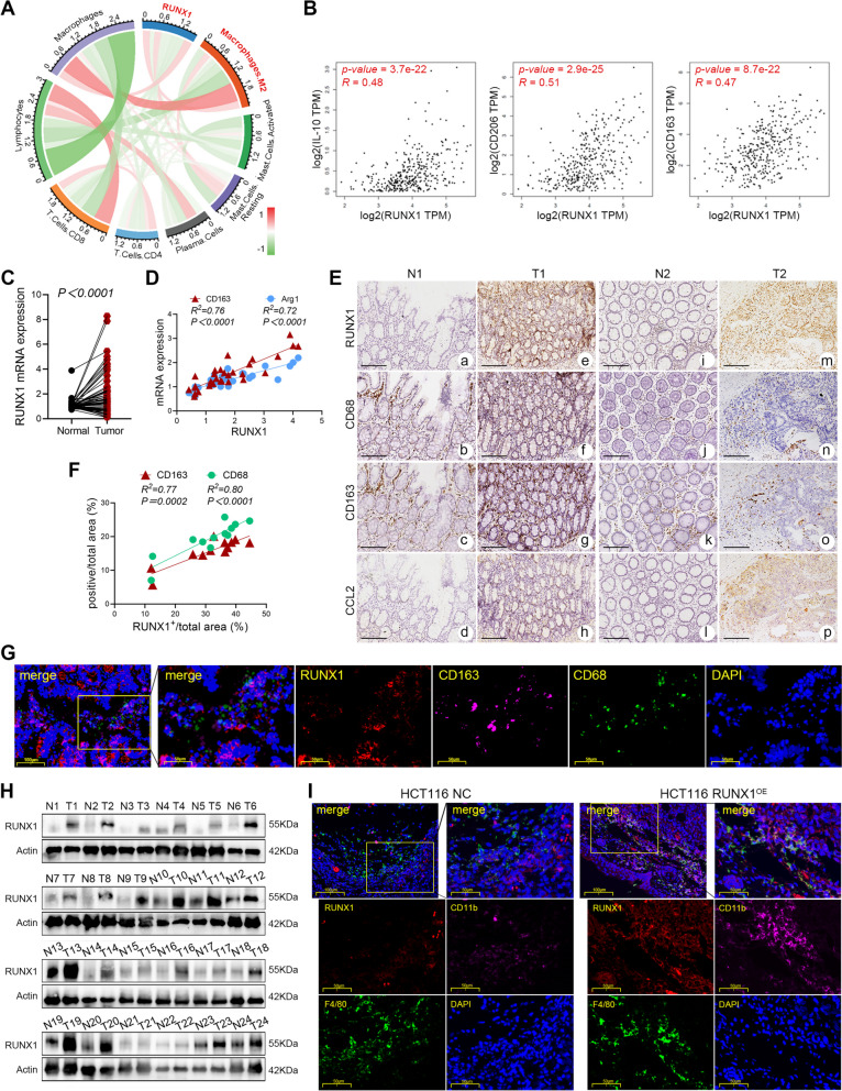
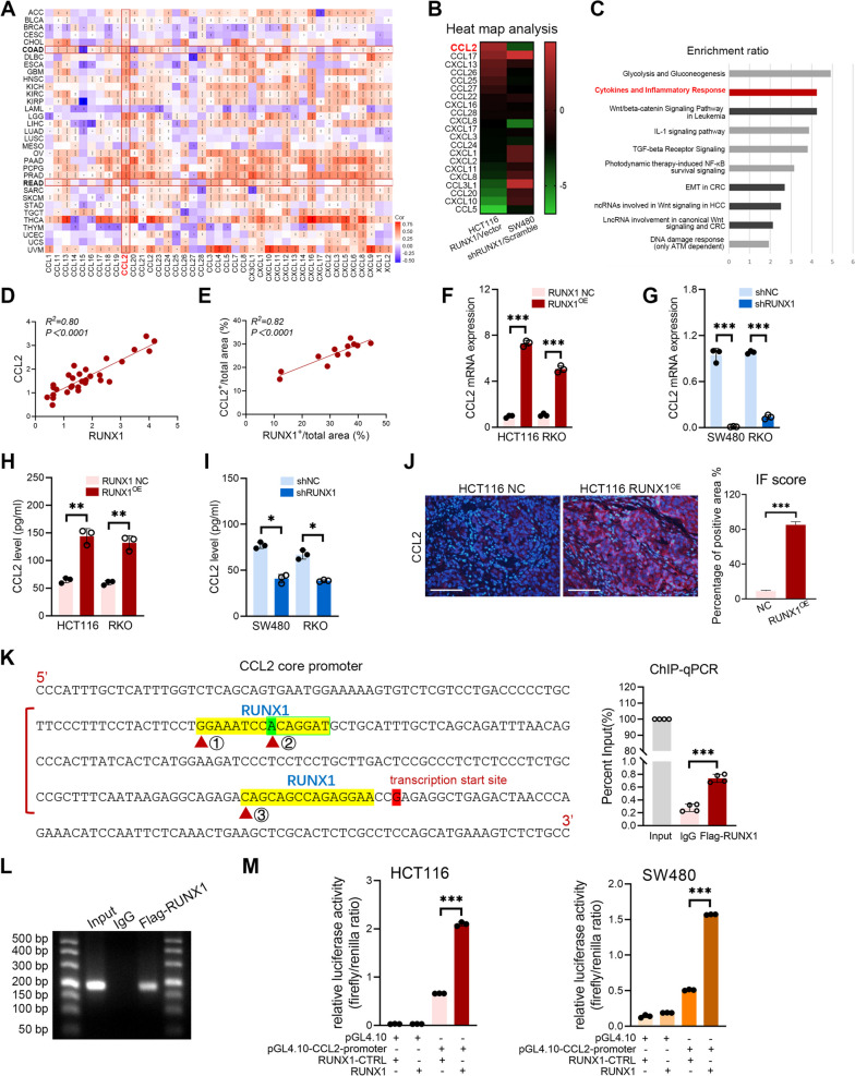
Colorectal cancer (CRC) is a common malignancy worldwide. Angiogenesis and metastasis are the critical hallmarks of malignant tumor. Runt-related transcription factor 1 (RUNX1), an efficient transcription factor, facilitates CRC proliferation, metastasis and chemotherapy resistance. We aimed to investigate the RUNX1 mediated crosstalk between tumor cells and M2 polarized tumor associated macrophages (TAMs) in CRC, as well as its relationship with neoplastic angiogenesis. We found that RUNX1 recruited macrophages and induced M2 polarized TAMs in CRC by promoting the production of chemokine 2 (CCL2) and the activation of Hedgehog pathway. In addition, we found that the M2 macrophage-specific generated cytokine, platelet-derived growth factor (PDGF)-BB, promoted vessel formation both in vitro and vivo. PDGF-BB was also found to enhance the expression of RUNX1 in CRC cell lines, and promote its migration and invasion in vitro. A positive feedback loop of RUNX1 and PDGF-BB was thus formed. In conclusion, our data suggest that RUNX1 promotes CRC angiogenesis by regulating M2 macrophages during the complex crosstalk between tumor cells and TAMs. This observation provides a potential combined therapy strategy targeting RUNX1 and TAMs-related PDGF-BB in CRC.

Keywords: Angiogenesis; Colorectal cancer; M2 polarization; RUNX1; Tumor associated macrophages.


Conflict of interest statement

The authors declare no competing interests.

Figures

Fig. 1
Fig. 1
RUNX1 promotes TAMs infiltration in CRC. A Circular plot of immune cells enriched for the RUNX1-associated genes in COAD. RUNX1 expression is well related to M2 macrophage in COAD. B Correlation analysis between RUNX1 and IL-10 (left), CD206 (middle) and CD163 (right) in the TCGA database. C RT-qPCR analysis of the RUNX1 mRNA expression in tumor specimens and matched normal tissues (n = 66). D Correlation between the mRNA expression of RUNX1 and CD163, Arg1 in CRC tissues (n = 30). E Representative IHC analysis of RUNX1, CD68, CD163 and CCL2 expression in 12 pairs of cancer and the adjacent normal tissue from CRC patients. Scale bars: 100 μm. F Correlation between the expression of RUNX1 and CD68, CD163 in the IHC analysis. G Multi-label immunofluorescence staining of tumor tissue in patients with CRC. H The expression of RUNX1 in CRC tumor tissues and matched normal tissues was determined by western blotting (n = 24). I Multi-label immunofluorescence staining of tumor tissue from the nude mouse orthotopic CRC tumors
Fig. 2
Fig. 2
RUNX1 promotes CCL2-mediated recruitment of TAMs. A Correlation between the RUNX1 expression and chemokine family members in 33 TCGA tumors. B Heat map analysis of the chemokines and its receptors based on the RNA-Seq data of HCT116 RUNX1OE/vector and SW480 shRUNX1/scramble. C Enrichment analysis of RUNX1-related signaling pathways using Wikipathway Cancer platform. D Correlation analysis between the RUNX1 mRNA and CCL2 mRNA in tumor specimens (n = 30). E Correlation analysis between RUNX1 IHC staining and CCL2 staining in 12 pairs of CRC samples. F, G RT-qPCR analysis of the expression of CCL2 mRNA in stably transfected CRC cells. H, I ELISA analysis of the CCL2 generation in supernatant of RUNX1 over-expressed or knockdown CRC cells. J (left) Representative IF staining of CCL2 in the orthotopic CRC tumors with RUNX1 over-expressed. Scale bars: 200 μm. (right) Quantification of percentage of CCL2+ HCT116 cells (of HCT116 cells; n = 3). Quantitative data are indicated as mean ± SEM. K (left) Possible binding sites of RUNX1 in the CCL2 core promoter. (right) ChIP-qPCR assay in HCT116. L All PCR products were confirmed by DNA agarose gel electrophoresis. M Dual-Glo luciferase assay in HCT116 and SW480
Fig. 3
Fig. 3
RUNX1 regulates M2 polarization of TAMs. A Experimental scheme of different CMs preparation and in vitro model of cells co-culture. B THP-1 was stimulated with CMs derived from RUNX1 over-expressed (left; HCT116 vector/RUNX1OE, RKO vector/RUNX1OE) or knockdown (right; SW480 shNC/shRUNX1, RKO shNC/shRUNX1) CRC cells, and the IL-10 in supernatant was analyzed by ELISA. C (left) Flow cytometry analysis of macrophage polarization phenotype stimulated by CMs derived from RUNX1 over-expressed HCT116 and RKO cells. (right) Quantification of percentage of CD68+CD206+ or CD68+CD200R+ macrophages (of macrophages; n = 3). D RT-qPCR analysis of the expression of CD163 mRNA, CD206 mRNA, Arg1 mRNA and IL-10 mRNA in macrophages treated with CMs derived from RUNX1 over-expressed HCT116 (left) and RKO (right) cells. (E) (left) Flow cytometry analysis of macrophage polarization state stimulated by CMs derived from RUNX1 knockdown SW480 and RKO cells. (right) Quantification of percentage of CD68+CD206+ or CD68+CD200R+ macrophages (of macrophages; n = 3). (F) RT-qPCR analysis of the expression of CD163 mRNA, CD206 mRNA, Arg1 mRNA and IL-10 mRNA in macrophages treated with CMs derived from RUNX1 knockdown SW480 (left) and RKO (right) cells. G CD206 and CD200R IF labeling of THP-1 cells treated with CRC cells-derived CMs. Data were recorded by a confocal laser scanning microscopy. Scale bars: 10 μm. H Quantification of mean fluorescence intensity of CD206 and CD200R (IntDen/Area; n = 3). I The expression of PTCH1, PTCH2, GLI1, SUFU and Shh in TAMs treated with CMs derived from RUNX1 over-expressed HCT116 cells was determined by western blotting. J (left) Flow cytometry analysis of macrophage phenotype induced by CMs derived from RUNX1 over-expressed HCT116 cells in the presence or absence of GDC-0449. (right) Quantification of percentage of CD68+CD206+ or CD68+CD200R+ macrophages (of macrophages; n = 3)
Fig. 4
Fig. 4
M2 TAMs derived PDGF-BB promotes tumor angiogenesis in vitro. A Gene set enrichment analysis in COAD conducted on the TCGA database. B Correlation analysis between RUNX1 and endothelial cell markers in CRC tissue samples. C Correlation analysis between RUNX1 and PDGFB (left), PDGFRA (middle) and PDGFRB (right) in the TCGA database. D Wikipathway cancer over-respresentation analysis of RUNX1 associated pathways. E The expression of PDGF-BB in THP-1 treated with CMs derived from RUNX1 over-expressed HCT116 and RKO cells was determined by ELISA. F The expression of PDGF-BB in THP-1 treated with CMs derived from RUNX1 down-regulated SW480 and RKO cells was determined by ELISA. G The expression of PDGF-BB in HCT116 RUNX1OE/THP-1 co-culture supernatant was determined by ELISA. H The expression of PDGF-BB in SW480 shRUNX1/THP-1 co-culture supernatant was determined by ELISA. I The 48 h proliferation rate of HUVECs treated with HCT116 RUNX1OE/THP-1 co-culture supernatant with or without anti-hPDGF-BB antibody (20 ng/ml) was detected. J (left) Transwell migration assay. The ability of HCT116 RUNX1OE/THP-1 co-culture supernatant to promote HUVECs migration in the presence or absence of anti-hPDGF-BB antibody (20 ng/ml) was evaluated. Scale bars: 200 μm. (right) Quantification of the number of cells migrating to the lower chamber. K (left) Transwell invasion assay. The ability of HCT116 RUNX1OE/THP-1 co-culture supernatant to promote HUVECs invasion in the presence or absence of anti-hPDGF-BB antibody (20 ng/ml) was evaluated. Scale bars: 200 μm. (right) Quantification of the number of cells invading to the lower chamber. L (left) Tube formation on Matrigel. The ability of HCT116 RUNX1OE/THP-1 co-culture supernatant to promote HUVECs tube formation in the presence or absence of anti-hPDGF-BB antibody (20 ng/ml) was evaluated. Scale bars: 200 μm. (right) Quantification of the branch points of HUVECs. M (left) Transwell migration assay. The ability of PDGF-BB to promote HUVECs migration was evaluated. Scale bars: 200 μm. (right) Quantification of the number of cells migrating to the lower chamber. N (left) Transwell invasion assay. The ability of PDGF-BB to promote HUVECs invasion was evaluated. Scale bars: 200 μm. (right) Quantification of number of cells invading to the lower well. O (left) Tube formation on Matrigel. The ability of PDGF-BB to promote HUVECs tube formation was examined. Scale bars: 200 μm. (right) Quantification of the branch points of HUVECs
Fig. 5
Fig. 5
RUNX1 induces crosstalk between CRC cells and TAMs to promote tumor angiogenesis. A Correlation analysis of the expression of RUNX1 and CD31 in different stages of CRC tissues by IHC. B Representative H&E and CD31 IF analysis of orthotopic CRC tumors in nude mice. Scale bars: 200 μm. C The number of blood vessels in H&E sections of orthotopic tumor. (n = 4, P < 0.01). D In vivo CAM assay. Photographs of the CAM assay showing a 9th day fertilized egg subjected to PBS, HCT116 NC-derived CMs, HCT116 RUNX1OE-derived CMs, THP-1-derived CMs, HCT116 NC/THP-1 co-culture-derived CMs, HCT116 RUNX1OE/THP-1 co-culture-derived CMs. E The ratio of vascular area to CAM area was calculated using the software ImageJ. F (left) Matrigel plug assay in nude mice. Representative H&E (g-l) and CD31 IHC (m-r) staining analysis of the viscous plugs. Scale bars: 100 μm. (right) The number of blood vessels in H&E sections of orthotopic tumor (n = 3, P < 0.001)
Fig. 6
Fig. 6
M2-TAMs derived PDGF-BB promotes malignant biological behavior of CRC cells A The levels of PDGF-BB in THP-1 culture medium with or without IL-4 stimulation were detected by ELISA. B The RUNX1 expression in HCT116 and SW480 cells treated with M0 or M2 TAMs culture supernatant in the presence or absence of anti-hPDGF-BB antibody were detected by western blotting. C RT-qPCR analysis of the RUNX1 mRNA expression in CRC cells treated with M0 or M2 TAMs culture supernatant. D (left) Transwell invasion assay. The invasion rate of HCT116 and SW480 cells treated with M0 or M2 TAMs culture supernatant were evaluated. Scale bars: 200 μm. (right) Quantification of the number of cells invading to the lower well. E (left) Transwell migration assay. The migration rate of HCT116 and SW480 cells treated with M0 or M2 TAMs culture supernatant were evaluated. Scale bars: 200 μm. (right) Quantification of the number of cells migrating to the lower chamber. F Wound healing assay. The ability of the M0 or M2 TAMs culture supernatant to promote CRC cells wound healing was evaluated. G The changes of RUNX1 expression in the cytosol and nuclei of HCT116 and SW480 cells upon the M0 or M2 TAMs culture supernatant stimulation were assessed by western blotting. H The changes of RUNX1 expression in the cytosol and nuclei of CRC cells treated with PBS or 20 ng/ml PDGF-BB were detected by western blotting. I, J RUNX1 IF labeling of HCT116 or SW480 cells treated with M0 or M2 TAMs culture supernatant. Data were recorded by confocal laser scanning. Scale bars: 10 μm. K The protein expression of c-myc, LEF1, cyclinD1 and GAPDH in HCT116 and SW480 cells treated with PBS or 20 ng/ml PDGF-BB were detected by western blotting. L The pulmonary metastasis model of CRC in nude mice. (left, middle) The full image of metastatic tumors. HCT116 cells were firstly stimulated with THP-1-derived CMs for 24h, and then inoculated into nude mice via caudal vein. (right) Quantification of numbers of the lung tumor nodules in different groups (n = 5). M Representative H&E staining analysis of the lung tumor nodules (#3, #4). Scale bars: 100 μm
Fig. 7
Fig. 7
RUNX1 promotes angiogenesis in CRC by regulating the crosstalk between tumor cells and TAMs. Summary diagram showing a complex interaction in the crosstalk of CRC cells and TAMs. RUNX1 derived from CRC cells recruits macrophages and induces the M2 polarization phenotype. The latter contributes to tumor angiogenesis in vivo and in vitro and migration and invasion of CRC cells via PDGF-BB, so as to form a positive feedback loop to promote the progression of CRC

References

    1. Sung H, Ferlay J, Siegel RL, Laversanne M, Soerjomataram I, Jemal A, et al. Global cancer statistics 2020: GLOBOCAN estimates of incidence and mortality worldwide for 36 cancers in 185 countries. CA Cancer J Clin. 2021;71:209–249. doi: 10.3322/caac.21660. - DOI - PubMed
    1. Teleanu RI, Chircov C, Grumezescu AM, Teleanu DM. Tumor Angiogenesis and Anti-Angiogenic Strategies for Cancer Treatment. J Clin Med. 2019;9:84. doi: 10.3390/jcm9010084. - DOI - PMC - PubMed
    1. Lugano R, Ramachandran M, Dimberg A. Tumor angiogenesis: causes, consequences, challenges and opportunities. Cell Mol Life Sci. 2020;77:1745–1770. doi: 10.1007/s00018-019-03351-7. - DOI - PMC - PubMed
    1. Folkman J. Angiogenesis: an organizing principle for drug discovery? Nat Rev Drug Discov. 2007;6:273–286. doi: 10.1038/nrd2115. - DOI - PubMed
    1. Kircher SM, Nimeiri HS, Benson AB., 3rd Targeting Angiogenesis in Colorectal Cancer: Tyrosine Kinase Inhibitors. Cancer J. 2016;22:182–189. doi: 10.1097/PPO.0000000000000192. - DOI - PubMed

LinkOut - more resources