. 2024 Feb 28;22(1):3.
doi: 10.1186/s13053-024-00275-7.

Preoperative multimodal ultrasonic imaging in a case of Peutz-Jeghers syndrome complicated by atypical lobular endocervical glandular hyperplasia: a case report and literature review

Affiliations

Preoperative multimodal ultrasonic imaging in a case of Peutz-Jeghers syndrome complicated by atypical lobular endocervical glandular hyperplasia: a case report and literature review

Liwen Yang et al. Hered Cancer Clin Pract. .

Abstract

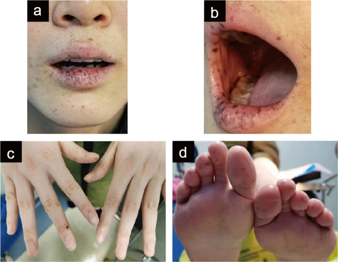
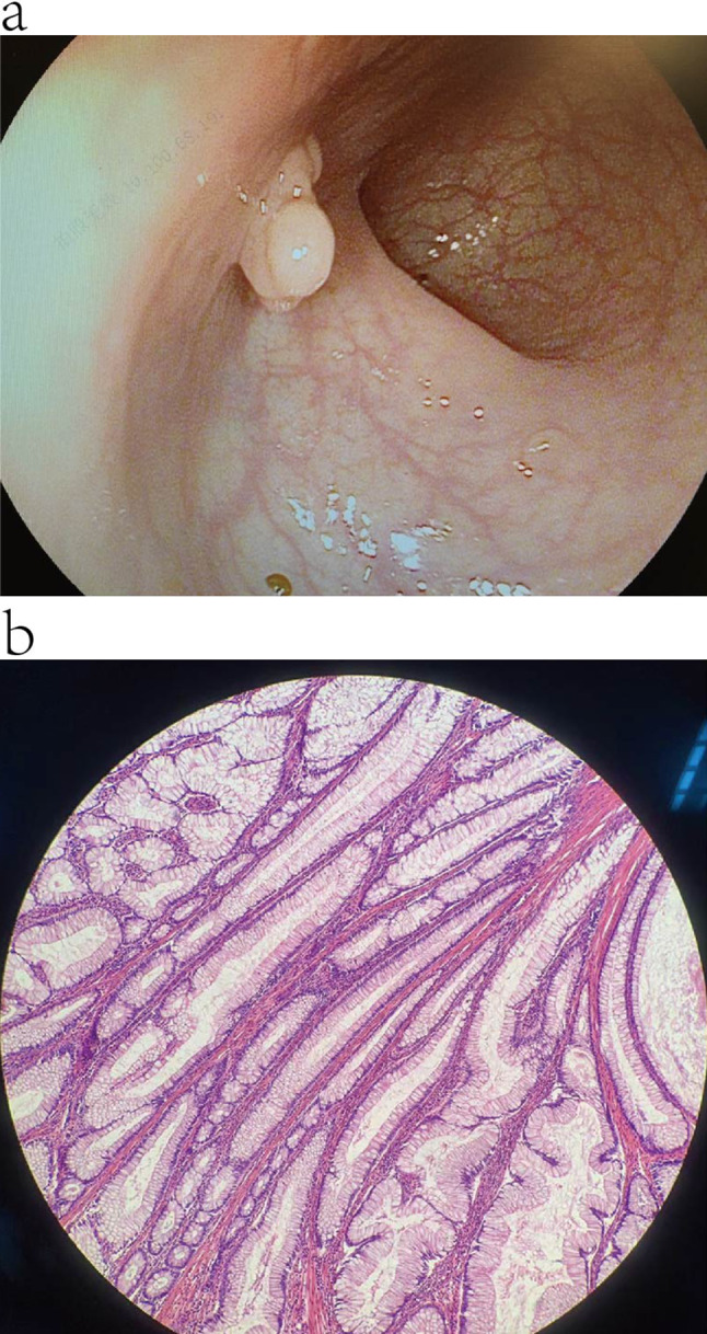
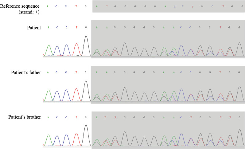
Background: Peutz-Jeghers syndrome (PJS), an autosomal dominant multiple cancerous disorder, is clinically characterized by mucocutaneous macules and multiple gastrointestinal hamartomatous polyps. Gastric-type endocervical adenocarcinoma (G-EAC), a special subtype of cervical adenocarcinoma with non-specific symptoms and signs, is known to occur in approximately 11% of female patients with PJS.

Case presentation: Here, we report a case of PJS in a 24-year-old female with multiple mucocutaneous black macules who complained of vaginal discharge and menorrhagia. Moreover, we first described the multimodal ultrasonographical manifestations of PJS-correlated G-EAC. The three-dimensional reconstructed view of G-EAC on 3D realisticVue exhibited a distinctive "cosmos pattern" resembling features on magnetic resonance imaging, and the contrast-enhanced ultrasound displayed a "quick-up and slow-down" pattern of the solid components inside the mixed cervical echoes. We reported the multimodal ultrasonographical characteristics of a case of PJS-related G-EAC, as well as reviewed PJS-related literature and medical imaging features and clinical characteristics of G-EAC to provide insight into the feasibility and potential of utilizing multimodal ultrasonography for the diagnosis of G-EAC.

Conclusions: Multimodal ultrasound can visualize morphological features, solid components inside, and blood supplies of the G-EAC lesion and distinguish the G-EAC lesion from normal adjacent tissues. This facilitates preoperative diagnosis and staging of PJS-related G-EAC, thereby aiding subsequent health and reproductive management for patients with PJS.

We reported multimodal ultrasonographical characteristics of a case of Peutz-Jeghers syndrome-related gastric-type endocervical adenocarcinoma (G-EAC), indicating the potential use of multimodal ultrasonography for G-EAC diagnosis.

Keywords: Atypical lobular endocervical glandular hyperplasia; Contrast-enhanced ultrasonography; Gastric-type endocervical adenocarcinoma; Multimodal ultrasonography; Peutz-Jeghers syndrome; Three-dimensional ultrasonography.


Conflict of interest statement

The authors declare that they have no competing interests.

Figures

Fig. 1
Fig. 1
Multiple hyperpigmented macules on the lips (a), buccal mucosa (b), fingertips (c), and toes (d) of the patient
Fig. 2
Fig. 2
Polyp-like neoplasms in patient’s gastrointestinal track. (a) Gastrointestinal endoscopy reveals multiple polyp-like neoplasms. (b) histopathological examination of polyp resected from the mid-ileum
Fig. 3
Fig. 3
MRI examination of cervical lesions. (a) Cross-sectional plane on the T2WI fat suppression sequence exhibits an enlarged cervix with multilocular space-occupying lesions of various sizes abutted through irregular septae. (b) The sagittal plane of contrast-enhanced MRI reveals the noticeably enhanced cyst walls and septae. Abbreviation MRI, magnetic resonance imaging
Fig. 4
Fig. 4
Three-dimensional and contrast-enhanced ultrasonography imaging. (a) Three-dimensional view on 3D realisticVue (Samsung Medical, Seoul, South Korea) shows multilocular signs resembling the “cosmos pattern” in MRI. (b) Three-dimensional reconstructed coronary section of uterus and cervix presented by the CrystalVue software (Samsung Medical, Seoul, South Korea). The enlarged barrel-shaped cervix with multilocular resonances and an uninvaded uterine cavity can be observed. (c) CEUS (PHILIPS EPIQ7, Philips Healthcare, Seattle, WA, the United States) examination on the blood perfusion of the cervical lesion. The ROI and curve in blue indicate the perfused region and TIC of the solid component in the cervix; the ROI and curve in orange represent the perfused region and TIC of the myometrium. The solid components of the cervical mixed echo began developing at the 7th second and peaked at the 22nd second, presenting a pattern of “quick-up and slow-down.” (d) Three-dimensional power Doppler (General Electric Company, Boston, Massachusetts, the United States) displays the increased blood supplies with irregular ramification, part of which multiplies and forms a clump-like pattern. Abbreviation CEUS, contrast-enhanced ultrasonography; CX, cervix; EN, endometrium; MRI, magnetic resonance imaging; ROI, region of interest; TIC: time-intensity curve
Fig. 5
Fig. 5
Multimodal ultrasonography of cervical lesions. (a) Transrectal gray-scale US (GE voluson E10 RIC5-9-D, General Electric Company, Boston, Massachusetts, the United States) showing an enlarged cervix with multiple cystic lesions of various sizes and a few solid components. The solid components are hyperechoic. The cystic lesions are irregular, honeycomb-shaped, and of various sizes and were encompassed by relatively larger cysts. (b) Transabdominal CEUS ((PHILIPS EPIQ7, Philips Healthcare, Seattle, Washington, the United States) showing perfusion of the solid components of the cervical lesions before the myometrium. (c) Transabdominal CEUS image showing an unevenly enhanced uterine myometrium, unenhanced cystic echoes, and equally enhanced solid components when peaked. (d) Measurement of the mass volume using VOCAL software. (e) Three-dimensional TUI (General Electric Company, Boston, Massachusetts, the United States) of blood flow in the cervical lesion indicates that its blood supply is located in the intercyst septate and solid components. (f) Biplane transrectal US sagittal view using Biplane Endocavity convex-linear array transducer (ELC13-4U, Mindray, Shenzhen, Guangdong, China) revealing the normal structure of the vagina without penetration of the cancerous lesion. (g) Biplane transrectal US sagittal view showing intact and smooth rims of the anterior and posterior vaginal fornices without thickening or penetration of the lesion. Abbreviation AVF, anterior vaginal fornix; AVW, anterior vaginal wall; AVW, anterior vaginal wall; BL, bladder; CC, cystic component; CE, cervical effusion; CEUS, contrast-enhanced ultrasonography; CX, cervix; HE, hyperecho; PB, perineal body; PVF, posterior vaginal fornix; PVW, posterior vaginal wall; PVW, posterior vaginal wall; R, rectum; RVS, rectovaginal septum; SC, solid component; TUI, tomographic ultrasound imaging; UM, uterine myometrium; US, ultrasound
Fig. 6
Fig. 6
Hysteroscopic views of the uterine cavity and cervix. (a) Presence of multiple polyp-like neoplasms in the uterine cavity. (b) Presence of multilocular lesions in the cervix
Fig. 7
Fig. 7
Sequencing results for STK11 in the patient and her family members. Whole-exome sequencing (patient) and Sanger sequencing (patient’s father and brother) confirmed the pathogenic heterogeneous mutation of the STK11 gene mapped to Chr19:1207063–1,207,063, NM_000455.5:exon1:.150dup (p.Met51Aspfs*112)
Fig. 8
Fig. 8
Macroscopic view of the resected tissues. (a-b) The enlarged cervix, measuring 6 cm × 5 cm × 4 cm, exhibits a barrel-like shape; (c) the inside view of the cervix presents multiple cystic cavities with smooth walls, which contain a large amount of water-like clean mucous fluid; (d) Macroscopic view of the resected left ovarian cyst measured 5 cm × 4 cm × 3 cm. The cyst exhibits a thin wall and watery fluid inside
Fig. 9
Fig. 9
Postoperative pathohistological examination of resected cervical tissue. (a) The glandular lesion invading the cervix and lower part of the uterus; (b) AB/PAS staining shows the neutral mucus inside of the gastric-type cells; (c) Positive staining of MUC6 by IHC, indicating the glandular epithelium was gastric-type; (d & e) the presence of LEGH in the cervix; (f) The presence of SGM and LEGH in the cervix. Abbreviation AB/PAS, Alcian-blue/Periodic acid Schiff reagent; IHC, immunohistochemistry; LEGH, lobular endocervical glandular hyperplasia; SGM, simple gastric metaplasia

Similar articles

Cited by

References

    1. Latchford AR, Clark SK. Gastrointestinal aspects of Peutz-Jeghers syndrome. Best Pract Res Clin Gastroenterol. 2022;58–59:101789. doi: 10.1016/j.bpg.2022.101789. - DOI - PubMed
    1. Wagner A, Aretz S, Auranen A, Bruno MJ, Cavestro GM, Crosbie EJ, et al. The management of Peutz-Jeghers syndrome: European hereditary tumour group (EHTG) guideline. J Clin Med. 2021;10. 10.3390/jcm10030473. - PMC - PubMed
    1. Hearle N, Schumacher V, Menko FH, Olschwang S, Boardman LA, Gille JJ, et al. Frequency and spectrum of cancers in the Peutz-Jeghers syndrome. Clin Cancer Res. 2006;12:3209–15. doi: 10.1158/1078-0432.CCR-06-0083. - DOI - PubMed
    1. Xu ZX, Jiang LX, Chen YR, Zhang YH, Zhang Z, Yu PF, et al. Clinical features, diagnosis, and treatment of Peutz-Jeghers syndrome: experience with 566 Chinese cases. World J Gastroenterol. 2023;29:1627–37. doi: 10.3748/wjg.v29.i10.1627. - DOI - PMC - PubMed
    1. Latchford AR, Phillips RKS. Gastrointestinal polyps and cancer in Peutz-Jeghers syndrome: clinical aspects. Fam Cancer. 2011;10:455–61. doi: 10.1007/s10689-011-9442-1. - DOI - PubMed

LinkOut - more resources