. 2024 Feb 28;19(1):34.
doi: 10.1186/s13020-024-00908-y.

The combination of Tanshinone IIA and Astragaloside IV attenuates myocardial ischemia-reperfusion injury by inhibiting the STING pathway

Affiliations

The combination of Tanshinone IIA and Astragaloside IV attenuates myocardial ischemia-reperfusion injury by inhibiting the STING pathway

Pan Zhai et al. Chin Med. .

Abstract

Background: Astragaloside IV (As-IV) and Tanshinone IIA (Ta-IIA) are the main ingredients of traditional Chinese medicinal Astragalus membranaceus (Fisch.) Bunge and Salvia miltiorrhiza Bunge, respectively, both of which have been employed in the treatment of cardiovascular diseases. Nevertheless, the efficacy of the combination (Co) of Ta-IIA and As-IV for cardiovascular diseases remain unclear and warrant further investigation. This study aimed to investigate the efficacy and the underlying molecular mechanism of Co in treating myocardial ischemia-reperfusion injury (MIRI).

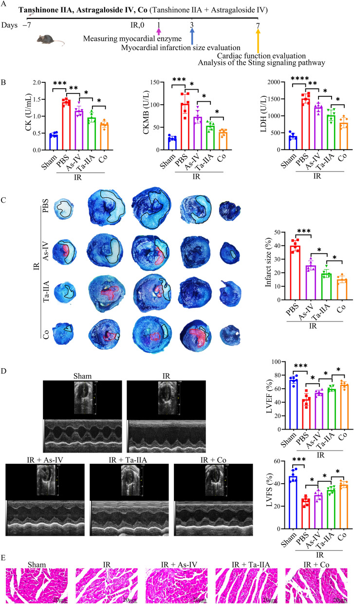
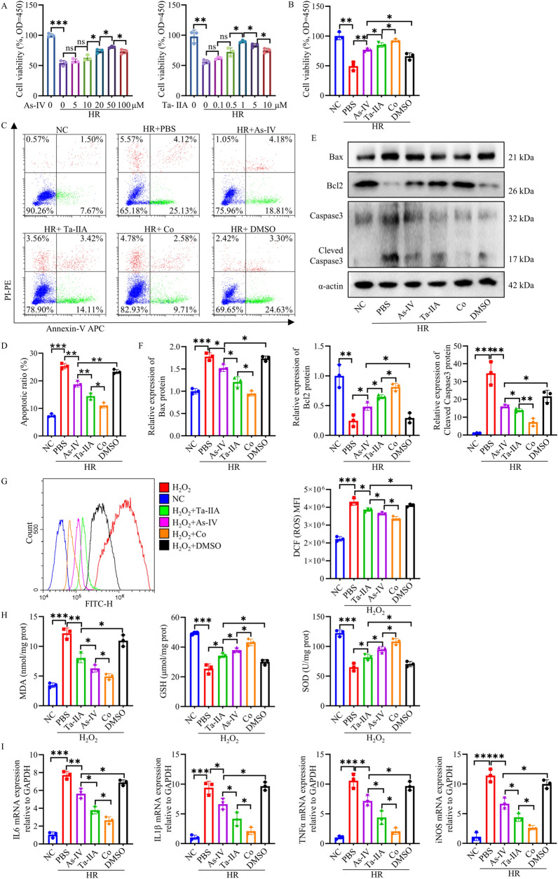
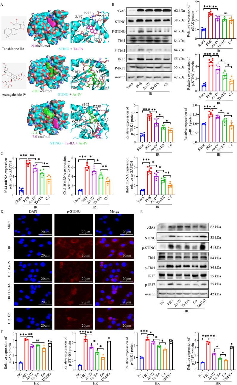
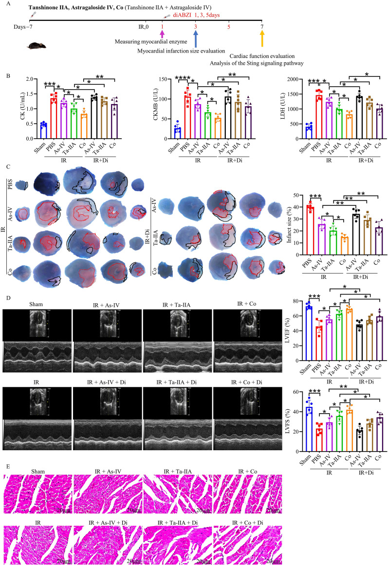
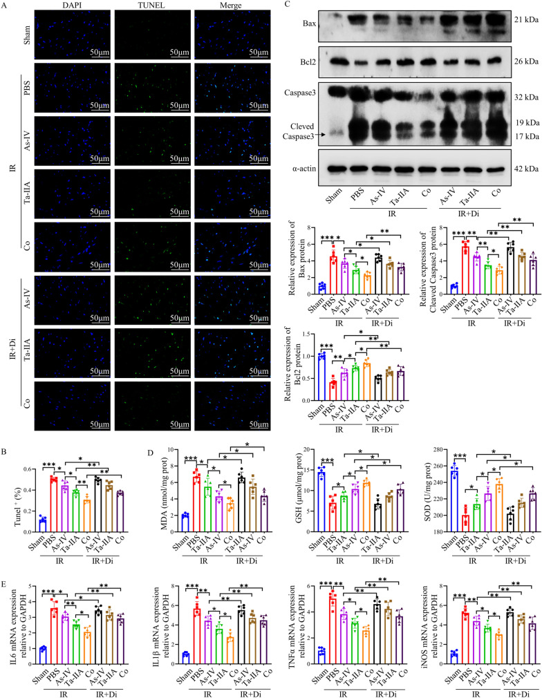
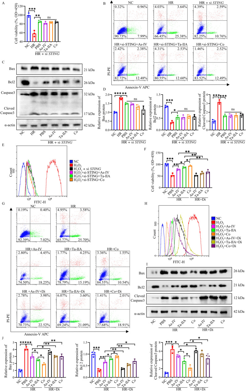
Methods: In order to assess the efficacy of Co, an in vivo MIRI mouse model was created by temporarily blocking the coronary arteries for 30 min and then releasing the blockage. Parameters such as blood myocardial enzymes, infarct size, and ventricular function were measured. Additionally, in vitro experiments were conducted using HL1 cells in both hypoxia-reoxygenation model and oxidative stress models. The apoptosis rate, expression levels of apoptosis-related proteins, oxidative stress indexes, and release of inflammatory factors were detected. Furthermore, molecular docking was applied to examine the binding properties of Ta-IIA and As-IV to STING, and western blotting was performed to analyze protein expression of the STING pathway. Additionally, the protective effect of Ta-IIA, As-IV and Co via inhibiting STING was further confirmed in models of knockdown STING by siRNA and adding STING agonist.

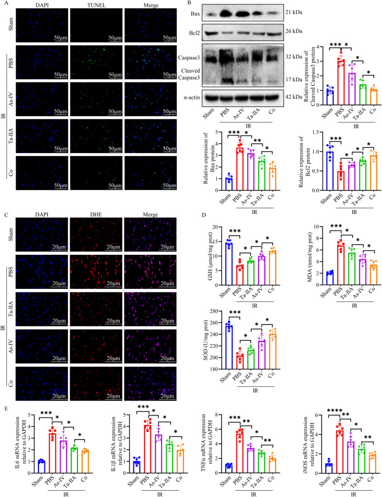
Results: Both in vitro and in vivo data demonstrated that, compared to Ta-IIA or As-IV alone, the Co exhibited superior efficacy in reducing the area of myocardial infarction, lowering myocardial enzyme levels, and promoting the recovery of myocardial contractility. Furthermore, the Co showed more potent anti-apoptosis, antioxidant, and anti-inflammation effects. Additionally, the Co enhanced the inhibitory effects of Ta-IIA and As-IV on STING phosphorylation and the activation of STING signaling pathway. However, the administration of a STING agonist attenuated the protective effects of the Co, Ta-IIA, and As-IV by compromising their anti-apoptotic, antioxidant, and anti-inflammatory effects in MIRI.

Conclusion: Compared to the individual administration of Ta-IIA or As-IV, the combined treatment demonstrated more potent ability in inhibiting apoptosis, oxidative stress, inflammation, and the STING signaling pathway in the context of MIRI, indicating a more powerful protective effect against MIRI.

Keywords: Apoptosis; Astragaloside IV; Myocardial ischemia reperfusion injury; Oxidative stress; STING pathway; Tanshinone IIA.


Conflict of interest statement

The authors declare that they have no competing interests.

Figures

Fig. 1
Fig. 1
The protective effect of As-IV, Ta-IIA, and Co on the heart of MIRI mice. Ta-IIA treatment (10 mg/kg/day, i.p.), As-IV treatment (15 mg/kg/day, i.p.), Co (Ta-IIA 10 mg/kg/day + As-IV 15 mg/kg/day i.p.) treatment were given 7 days before reperfusion until tested after anesthesia. Myocardial enzymes and LDH were detected 1 day after surgery, EB/TTC double staining was detected 3 days after surgery, and cardiac function was detected by echocardiography and HE staining 7 days after surgery. A Experimental design of As-IV, Ta-IIA, and Co for in vivo cardioprotection in mice. B Serum CK, CKMB levels, and serum LDH levels. C Representative digital images of heart sections by Evans blue and TTC double staining, and the percentage of infarct area. The blue-stained portion indicates the normal region, the red-stained portion indicates the ischemic region, and the white portion indicates the infarcted region. The ratio of the infarct area of the largest heart section to the total area of the section was chosen to be the percentage of infarct area. D Representative M-mode echocardiographic images, and the quantitative analysis of the LVEF and LVFS. E Representative picture of HE staining of myocardial tissue. Data are presented as mean ± SEM. (n = 6 in each group). *p < 0.05, **p < 0.01, ***p < 0.001
Fig. 2
Fig. 2
As-IV, Ta-IIA, and Co inhibited myocardial cell apoptosis, oxidative stress, and inflammatory response in MIRI. A Representative images of apoptotic cardiomyocytes. The apoptotic cells were detected by TUNEL (green), and the nuclei were detected by DAPI (blue). Scale bar = 50 μm. B Representative blots of the apoptosis-related proteins Bax, Bcl2, Caspase3, and Cleaved Caspase3 in myocardial tissue, as well as quantitative analysis of Bax, Bcl2, and Cleaved Caspase3. C Representative images of ROS content in cardiomyocytes. ROS was detected by DHE fluorescence staining (red), and nuclei were detected by DAPI (blue). scale bar = 20 μm. D Myocardial glutathione (GSH) activity, malondialdehyde(MDA) content, superoxide dismutase (SOD) activity. E. The mRNA quantification of cytokines IL-6, IL-1β, TNFα, iNOS in cardiomyocardial tissues. Data are presented as mean ± SEM. (n = 6 in each group). *p < 0.05, **p < 0.01, ***p < 0.001
Fig. 3
Fig. 3
Effects of As-IV and Ta-IIA on HR-induced apoptosis, oxidative stress, and inflammation in HL1 cells. A As-IV and Ta-IIA treatment dose- dependently increased the viability of HR- injured HL1 cells. B The increase in cell activity with Co treatment was greater in HR-injured HL1 cells than in those treated with Ta-IIA and As-IV. Ta-IIA was 1 μm, and As-IV was 50 μm. Co was a combination of Ta-IIA1 μm and As-IV 50 μm. C. Representative results of apoptosis rates measured in HL1 cells using flow cytometry. D Quantitative analysis of the apoptosis rate in (C). E Representative western blots of Bax, Bcl2, Cleaved Caspase3. F. The quantitative analysis of protein expression in (E). G Representative picture of the fluorescence curve of DCF in HL1 cells and the quantitative analysis of DCF mean fluorescence intensity. This was detected by flow cytometry. H. Cellular MDA content, GSH activity, SOD activity. I. Quantitative analysis of cytokines IL-6, IL-1β, TNFα and iNOS mRNA in HL1 cells. Data are presented as mean ± SEM. (n = 3 in each group). *p < 0.05, **p < 0.01, ***p < 0.001, ns = not statistically significant
Fig. 4
Fig. 4
As-IV and Ta-IIA inhibited the STING pathway. A Chemical structure and representative simulations of the binding to the STING protein for As-IV and Ta-IIA. B Representative western blots and quantification of STING pathway-associated proteins in myocardial tissue. C Quantification of mRNA expression of the cytokines Ifi44, Cxcl10 and Ifnb1 downstream of the STING pathway caused by MIRI. D Representative immunofluorescence staining of p-STING in HL1 cells. The p-STING proteins were stained red, and the nuclei were detected by DAPI (blue). Scale bar = 20 μm. E Representative western blots of STING pathway-associated proteins in HR-induced HL1 cells. F The quantitative analysis of protein expression in (E). Data are presented as mean ± SEM. (n = 6 in each group in vivo; n = 3 in each group in vitro). *p < 0.05, **p < 0.01, ***p < 0.001, ns = not statistically significant
Fig. 5
Fig. 5
STING agonist treatment attenuated the myocardial protection of Ta-IIA, As-IV, and Co on MIRI mice. Ta-IIA treatment (10 mg/kg/d, i.p.), As-IV treatment (15 mg/kg/day, i.p.), Co (Ta-IIA 10 mg/kg/day + As-IV 15 mg/kg/day i.p.) treatment were given 7 days before reperfusion until tested after anesthesia. DiabZI (3 mg/kg, i.v.) was administered once every other day for a total of three times following reperfusion surgery. A Experimental procedures for As-IV, Ta-IIA and Co in mice after the addition of agonists. B Serum CK, CKMB levels, and serum LDH levels. C Representative digital images of heart sections by Evans blue and TTC double staining, and the percentage of infarct area. The blue-stained portion indicates the normal region, the red-stained portion indicates the ischemic region, and the white portion indicates the infarcted region. The ratio of the infarct area of the largest heart section to the total area of the section was chosen to be the percentage of infarct area. D Representative M-mode echocardiographic images, and the quantitative analysis of the LVEF and LVFS. E Representative picture of HE staining of myocardial tissue. Data are presented as mean ± SEM. (n = 6 in each group). *p < 0.05, **p < 0.01, ***p < 0.001
Fig. 6
Fig. 6
The anti-apoptotic, antioxidant and anti-inflammatory effects of Ta-IIA, As-IV and Co were weakened by diabZI. A Representative images of apoptotic cardiomyocytes. The apoptotic cells were detected by TUNEL (green), and the nuclei were detected by DAPI (blue). Scale bar = 50 μm. B The quantitative analysis of Tunel+ cells of (A). C Representative blots of the apoptosis-related proteins in myocardial tissue, as well as quantitative analysis of Bax, Bcl2, and Cleaved Caspase3. D Myocardial MDA content, GSH activity, SOD activity. E. The mRNA quantification of cytokines IL-6, IL-1β, TNFα, iNOS in cardiomyocardial tissues. Data are presented as mean ± SEM. (n = 6 in each group). *p < 0.05, **p < 0.01, ***p < 0.001
Fig. 7
Fig. 7
The STING pathway is critical for Ta-IIA, As-IV, and Co effect on HR-induced HL1 cells. The effects of Ta-IIA, As-IV and Co on cell activity, apoptosis, oxidative stress and inflammation were detected in HL1 cells after STING siRNA and diabZI administration, respectively. A HL1 cells STING siRNA cell activity after Ta-IIA, As-IV and Co treatment. B Representative results of apoptosis rates measured in HL1 cells using flow cytometry. C. Representative western blots of Bax, Bcl2, Cleaved Caspase3. D The quantitative analysis of protein expression in (C). E Representative picture of the fluorescence curve of DCF in HL1 cells. F Cell activity of Ta-IIA, As-IV and Co treatment after diabZI administration. G Representative pictures of apoptosis rates of HL1 cells measured by flow cytometry. H Representative diagram of the DCF fluorescence curve in HL1 cells. I Representative western blotting of apoptosis-associated proteins. J Quantitative analysis of protein expression in (I). Data are presented as mean ± SEM. (n = 3 in each group). *p < 0.05, **p < 0.01, ***p < 0.001, ns = not statistically significant

Similar articles

Cited by

References

    1. Oseran AS, Yeh RW. Time to treatment in ST-segment elevation myocardial infarction: identifying dangerous delays or diminishing returns? JAMA. 2022;328(20):2016–2017. doi: 10.1001/jama.2022.19441. - DOI - PubMed
    1. Chen J, Huang Q, Li J, et al. Panax ginseng against myocardial ischemia/reperfusion injury: a review of preclinical evidence and potential mechanisms. J Ethnopharmacol. 2023;300:115715. doi: 10.1016/j.jep.2022.115715. - DOI - PubMed
    1. Schafer A, Konig T, Bauersachs J, et al. Novel therapeutic strategies to reduce reperfusion injury after acute myocardial infarction. Curr Probl Cardiol. 2022;47(12):101398. doi: 10.1016/j.cpcardiol.2022.101398. - DOI - PubMed
    1. Ibanez B, Heusch G, Ovize M, et al. Evolving therapies for myocardial ischemia/reperfusion injury. J Am Coll Cardiol. 2015;65(14):1454–1471. doi: 10.1016/j.jacc.2015.02.032. - DOI - PubMed
    1. Sun L, Wu J, Du F, et al. Cyclic GMP-AMP synthase is a cytosolic DNA sensor that activates the type I interferon pathway. Science. 2013;339(6121):786–791. doi: 10.1126/science.1232458. - DOI - PMC - PubMed