

Neurovascular anatomy of the developing human fetal penis and clitoris
- PMID: 38419143
- PMCID: PMC11161816
- DOI: 10.1111/joa.14029
Neurovascular anatomy of the developing human fetal penis and clitoris
Abstract
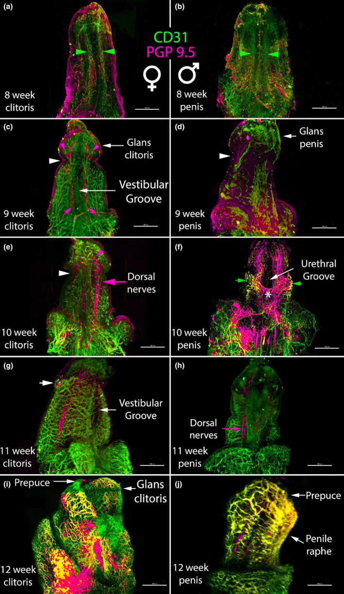
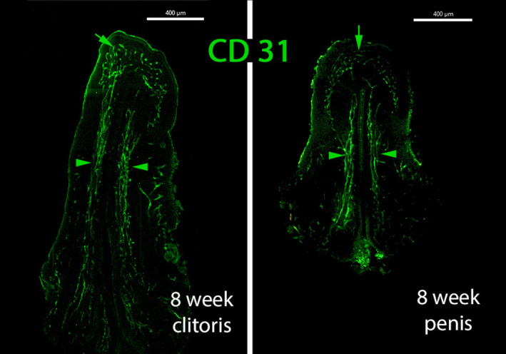
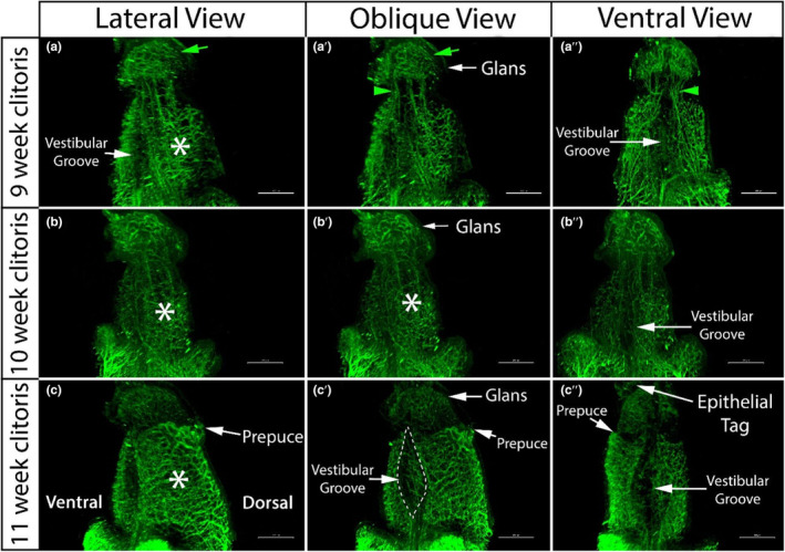
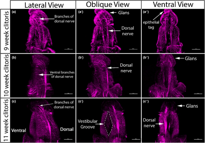
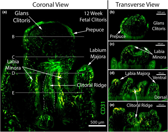
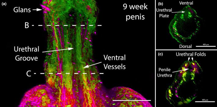
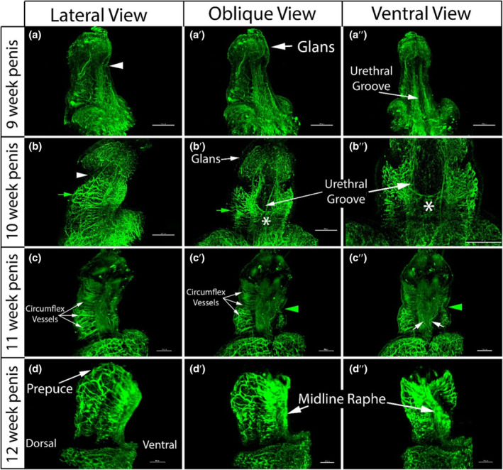
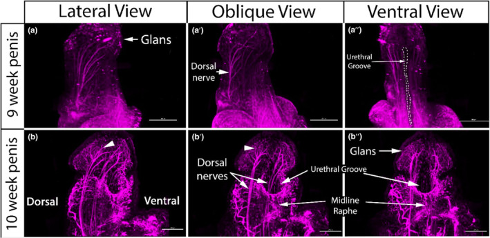
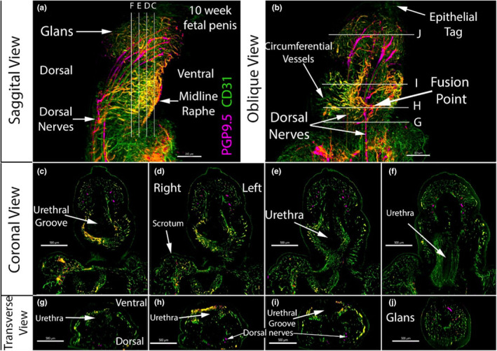
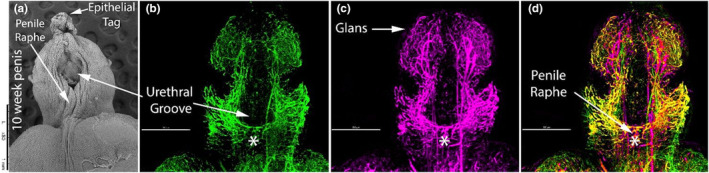
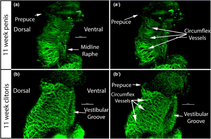
The human penile and clitoral development begins from a morphologically indifferent genital tubercle. Under the influence of androgen, the genital tubercle forms the penis by forming a tubular urethra within the penile shaft. Without the effect of the androgen, the genital tubercle differentiates into the clitoris, and a lack of formation of the urethra within the clitoris is observed. Even though there are similarities during the development of the glans penis and glans clitoris, the complex canalization occurring along the penile shaft eventually leads to a morphological difference between the penis and clitoris. Based on the morphological differences, the main goal of this study was to define the vascular and neuronal anatomy of the developing penis and clitoris between 8 and 12 weeks of gestation using laser scanning confocal microscopy. Our results demonstrated there is a co-expression of CD31, which is an endothelial cell marker, and PGP9.5, which is a neuronal marker in the penis where the fusion is actively occurring at the ventral shaft. We also identified a unique anatomical structure for the first time, the clitoral ridge, which is a fetal structure running along the clitoral shaft in the vestibular groove. Contrary to previous anatomical findings which indicate that the neurovascular distribution in the developing penis and clitoris is similar, in this study, laser scanning confocal microscopy enabled us to demonstrate finer differences in the neurovascular anatomy between the penis and clitoris.
Keywords: anatomy; clitoris; fetal; neurovascular; penis.
© 2024 The Authors. Journal of Anatomy published by John Wiley & Sons Ltd on behalf of Anatomical Society.
Conflict of interest statement
We have no conflicts of interest to declare.
Figures
Similar articles
-
Androgen and estrogen receptor expression in the developing human penis and clitoris.Differentiation. 2020 Jan-Feb;111:41-59. doi: 10.1016/j.diff.2019.08.005. Epub 2019 Sep 6. Differentiation. 2020. PMID: 31655443 Free PMC article.
-
Hot spots in fetal human penile and clitoral development.Differentiation. 2020 Mar-Apr;112:27-38. doi: 10.1016/j.diff.2019.11.001. Epub 2019 Dec 4. Differentiation. 2020. PMID: 31874420
-
Development of the human penis and clitoris.Differentiation. 2018 Sep-Oct;103:74-85. doi: 10.1016/j.diff.2018.08.001. Epub 2018 Aug 23. Differentiation. 2018. PMID: 30249413 Free PMC article.
-
Anatomy of the clitoris.J Urol. 2005 Oct;174(4 Pt 1):1189-95. doi: 10.1097/01.ju.0000173639.38898.cd. J Urol. 2005. PMID: 16145367 Review.
-
Anatomy of the clitoris and its impact on neophalloplasty (metoidioplasty) in female transgenders.Clin Anat. 2015 Apr;28(3):368-75. doi: 10.1002/ca.22525. Epub 2015 Mar 4. Clin Anat. 2015. PMID: 25740576 Review.
References
-
- Ban, S. , Sata, F. , Kurahashi, N. , Kasai, S. , Moriya, K. , Kakizaki, H. et al. (2008) Genetic polymorphisms of ESR1 and ESR2 that may influence estrogen activity and the risk of hypospadias. Human Reproduction, 23, 1466–1471. - PubMed
-
- Baskin, L. , Derpinghaus, A. , Cao, M. , Sinclair, A. , Li, Y. , Overland, M. et al. (2020) Hot spots in fetal human penile and clitoral development. Differentiation, 112, 27–38. - PubMed
-
- Baskin, L.S. , Erol, A.L.I. , Li, Y.W. & Cunha, G.R. (1998) Anatomical studies of hypospadias. The Journal of Urology, 160, 1108–1115. - PubMed
Publication types
MeSH terms
Grants and funding
LinkOut - more resources
Full Text Sources
