

Forward viewing liner echoendoscopy for therapeutic interventions
- PMID: 38419166
- PMCID: PMC10984744
- DOI: 10.5946/ce.2023.271
Forward viewing liner echoendoscopy for therapeutic interventions
Abstract
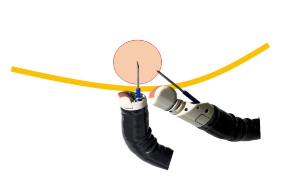
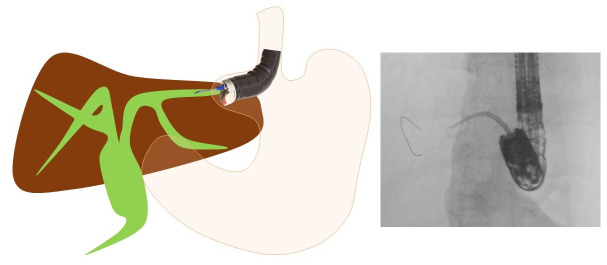
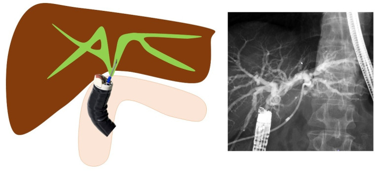
Therapeutic endoscopic ultrasonography (EUS) procedures using the forward-viewing convex EUS (FV-EUS) have been reviewed based on the articles reported to date. The earliest reported procedure is the drainage of pancreatic pseudocysts using FV-EUS. However, the study on drainage of pancreatic pseudocysts focused on showing that drainage is possible with FV-EUS rather than leveraging its features. Subsequently, studies describing the characteristics of FV-EUS have been reported. By using FV-EUS in EUS-guided choledochoduodenostomy, double punctures in the gastrointestinal tract can be avoided. In postoperative modified anatomical cases, using the endoscopic function of FV-EUS, procedures such as bile duct drainage from anastomosis, pancreatic duct drainage from the afferent limb, and abscess drainage from the digestive tract have been reported. When a perpendicular puncture to the gastrointestinal tract is required or when there is a need to insert the endoscope deep into the gastrointestinal tract, FV-EUS is considered among the options.
Keywords: Endoscopic ultrasonography-guided biliary drainage; Endosonography; Forward-viewing endoscopic ultrasonography; Interventional endoscopic ultrasonography; Therapeutic endoscopic ultrasonography.
Conflict of interest statement
The authors have no potential conflicts of interest.
Figures


References
-
- Kida M, Araki M, Miyazawa S, et al. Fine needle aspiration using forward-viewing endoscopic ultrasonography. Endoscopy. 2011;43:796–801. - PubMed
-
- Hara K, Yamao K, Hijioka S, et al. Prospective clinical study of endoscopic ultrasound-guided choledochoduodenostomy with direct metallic stent placement using a forward-viewing echoendoscope. Endoscopy. 2013;45:392–396. - PubMed
-
- Matsumoto S, Hara K, Mizuno N, et al. Risk factor analysis for adverse events and stent dysfunction of endoscopic ultrasound-guided choledochoduodenostomy. Dig Endosc. 2020;32:957–966. - PubMed
Publication types
LinkOut - more resources
Full Text Sources
