. 2024 Jul;245(1):70-83.
doi: 10.1111/joa.14033. Epub 2024 Feb 28.

Effect of deletion of the protein kinase PRKD1 on development of the mouse embryonic heart

Affiliations

Effect of deletion of the protein kinase PRKD1 on development of the mouse embryonic heart

Qazi Waheed-Ullah et al. J Anat. 2024 Jul.

Abstract

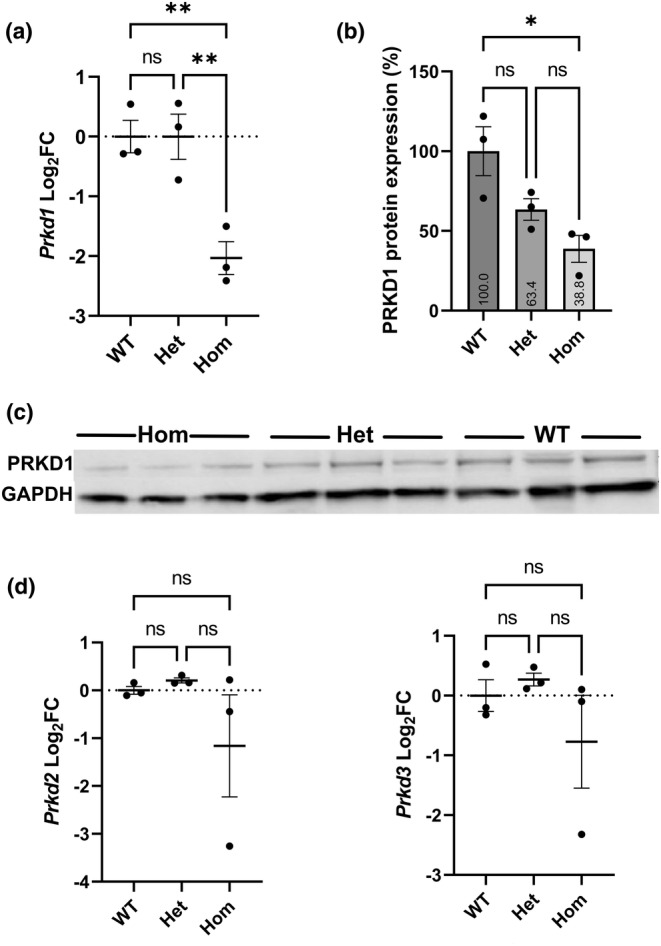
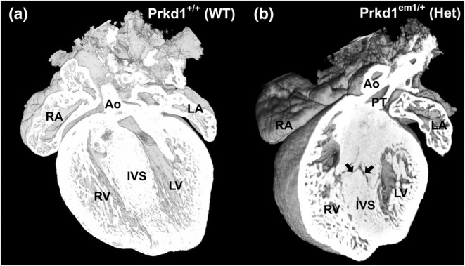
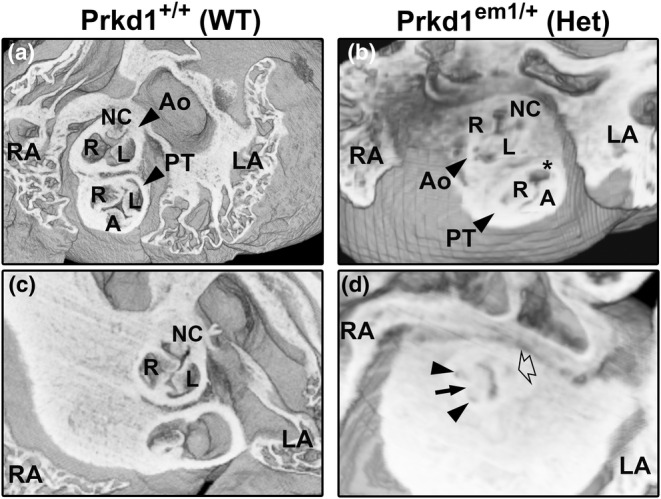
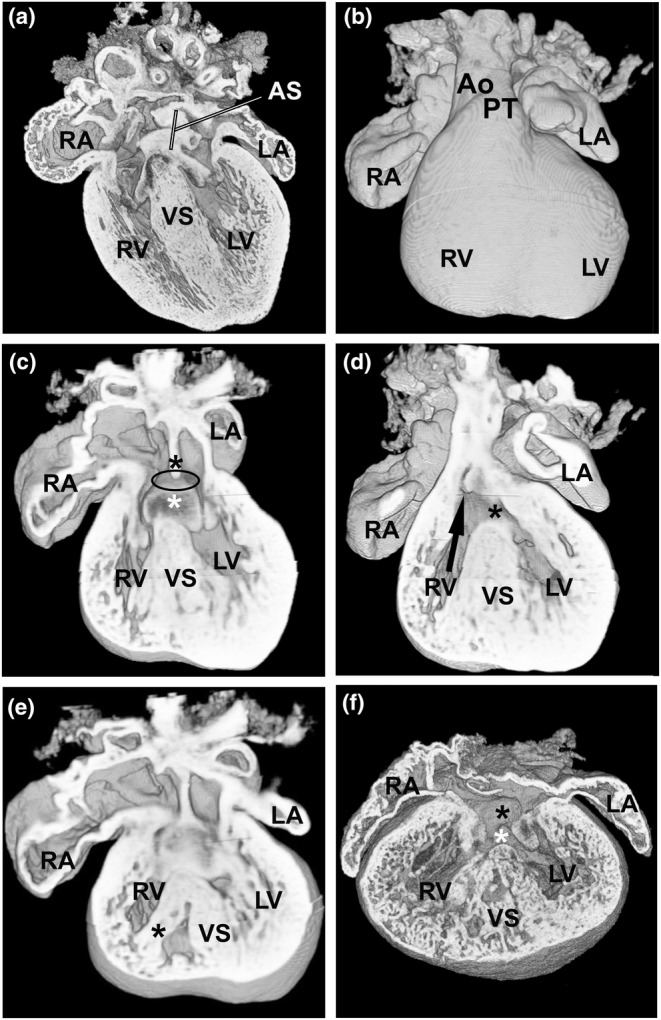
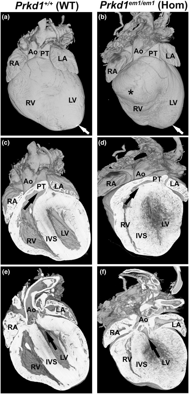
Congenital heart disease (CHD) is the most common congenital anomaly, with an overall incidence of approximately 1% in the United Kingdom. Exome sequencing in large CHD cohorts has been performed to provide insights into the genetic aetiology of CHD. This includes a study of 1891 probands by our group in collaboration with others, which identified three novel genes-CDK13, PRKD1, and CHD4, in patients with syndromic CHD. PRKD1 encodes a serine/threonine protein kinase, which is important in a variety of fundamental cellular functions. Individuals with a heterozygous mutation in PRKD1 may have facial dysmorphism, ectodermal dysplasia and may have CHDs such as pulmonary stenosis, atrioventricular septal defects, coarctation of the aorta and bicuspid aortic valve. To obtain a greater appreciation for the role that this essential protein kinase plays in cardiogenesis and CHD, we have analysed a Prkd1 transgenic mouse model (Prkd1em1) carrying deletion of exon 2, causing loss of function. High-resolution episcopic microscopy affords detailed morphological 3D analysis of the developing heart and provides evidence for an essential role of Prkd1 in both normal cardiac development and CHD. We show that homozygous deletion of Prkd1 is associated with complex forms of CHD such as atrioventricular septal defects, and bicuspid aortic and pulmonary valves, and is lethal. Even in heterozygotes, cardiac differences occur. However, given that 97% of Prkd1 heterozygous mice display normal heart development, it is likely that one normal allele is sufficient, with the defects seen most likely to represent sporadic events. Moreover, mRNA and protein expression levels were investigated by RT-qPCR and western immunoblotting, respectively. A significant reduction in Prkd1 mRNA levels was seen in homozygotes, but not heterozygotes, compared to WT littermates. While a trend towards lower PRKD1 protein expression was seen in the heterozygotes, the difference was only significant in the homozygotes. There was no compensation by the related Prkd2 and Prkd3 at transcript level, as evidenced by RT-qPCR. Overall, we demonstrate a vital role of Prkd1 in heart development and the aetiology of CHD.

Keywords: Prkd1; congenital heart disease; high‐resolution episcopic microscopy; protein kinase; protein kinase D1.


Conflict of interest statement

The authors declare they have no conflicts of interest.

Figures

FIGURE 1
FIGURE 1
Expression of Prkd1, Prkd2 and Prkd3 in E12.5 hearts. (a) Prkd1 mRNA expression in Prkd1 em1/+ (het) and Prkd1 em1/em1 (hom) hearts, compared to WT. A statistically significant difference in expression of Prkd1 in homozygous hearts compared to wild type (WT) (p = 0.009) and heterozygotes (p = 0.009) was found. (b) Mean normalized PRKD1 protein expression in percentage. Prkd1 em1/+ (het) and Prkd1 em1/em1 (hom) hearts were compared to WT (100%) using GAPDH as the reference protein. A significant difference was seen between homozygotes and WT controls (p = 0.017). (c) Representative western immunoblot of PRKD1 and GAPDH loading control in Prkd1 em1 E12.5 hearts. (d) Prkd2 and Prkd3 mRNA expressions in Prkd1 em1/+ (het) and Prkd1 em1/em1 (hom) hearts, compared to WT. No statistically significant difference in expression of Prkd2 or Prkd3 in homozygous hearts compared to WT (p = 0.43 and 0.52, respectively) and heterozygotes (p = 0.33 and 0.34, respectively) was found. For all experiments n = 3 per group, with each study repeated three times; error bars denote SEM; FC, fold change; n.s, not significant; **p < 0.01; *p < 0.1.
FIGURE 2
FIGURE 2
One heterozygous Prkd1 em1 E15.5 heart had a congenital heart defect. (a–b) Ventral view of a WT (a) and Prkd1 em1/ (b) heart. A small muscular ventricular septal defect can be seen in the Prkd1 em1/+ heart (arrows), in comparison to WT control where a normal interventricular septum (IVS) can be seen. The myocardial trabeculae also appear coarse and hypertrophic compared to controls. Ao, Aorta; LA, left atrium; LV, left ventricle; PT, pulmonary trunk; RA, right atrium; RV, right ventricle; WT, wild type.
FIGURE 3
FIGURE 3
Morphology of abnormal valves in outflow vessels in E15.5 Prkd1 em1/em1 (homozygous) hearts. (a, b) Superior views of a Prkd1 +/+ (a) and Prkd1 em1/em1 (b) heart. The WT heart has normal valves in the outflow region, with three valve leaflets (right, left and anterior) seen in the pulmonary trunk (a). In contrast, the homozygous heart has an abnormal pulmonary valve (b); a right and anterior leaflet could be discerned, but the left leaflet was deficient (asterisk), indicating a dysplastic leaflet in the pulmonary valve. Three leaflets (right, left and non‐coronary) can be seen in the aortic valve in both hearts. (c, d) A second E15.5 WT heart (c) and homozygous (d) heart from the superior aspect. The WT heart has normal leaflets (right, left and non‐coronary) in the aortic valve (c). In contrast the homozygous heart had an abnormal aortic valve (d). There are three sinuses (trisinuate; two are denoted by small arrows and one by an open arrow), but there were only two leaflets (bileaflet). The long black arrow points to the site of raphe, where the right and non‐coronary valve leaflets are fused. A, anterior; Ao, aorta; L, left; LA, left atrium; LV, left ventricle; NC, non‐coronary; PT, pulmonary trunk; R, right; RA, right atrium; RV, right ventricle; WT, wild type.
FIGURE 4
FIGURE 4
AVSD is seen in two E15.5 Prkd1 em1/em1 (homozygous) hearts. (a) A WT control heart with normal atrial septum. (b) An external ventral view of a Prkd1 em1/em1 heart with AVSD. (c) Same heart as in b; the lack of fusion of the dorsal endocardial cushion (white asterisk) with the septum primum (lack of fusion denoted by black oval) can be seen; the septum primum is indicated (black asterisk). The ventricular opening in this region is due to an inlet VSD. (d) A more ventral view shows the outlet VSD (black asterisk), with the aortic valve arising over the right ventricle (RV) (black arrow). The inlet and outlet VSDs are continuous. (e) A dorsal view, the right ventricle appears to be divided by either a large papillary muscle or a prominent septomarginal trabeculation (black asterisk). (f) Second homozygous heart with AVSD (black asterisk for atrial part and white asterisk for the inlet VSD). There is very marked trabeculation of the myocardium of both ventricles, with deep intertrabecular crypts and only a thin layer of compact myocardium present. There are a number of muscular openings within the ventricular septum. AS, atrial septum; Ao, aorta; LA, left atrium; LV, left ventricle; PT, pulmonary trunk; RA, right atrium; RV, right ventricle; IVS, interventricular septum; WT, wild type.
FIGURE 5
FIGURE 5
Bicuspid aortic valve is seen in P7 Prkd1 em1/+ heart. (a) Axial view of a Prkd1 +/+ (WT) heart at P7 stage showing a normal aortic valve with three sinuses of the three leaflets. (b) A Prkd1 em1/+ (Het) heart with a bicuspid aortic valve (AV), with two sinuses from the two leaflets (right and left) denoted by an asterisk (bisinuate and bileaflet). The mitral valve (MV) is also denoted, with normal appearing anterior (A) and posterior (P) leaflets. (b′) Inset in b is taken at a slightly different angle to show the right and left coronary ostia are patent (open arrows). R, right; L, left; NC, non‐coronary; Ao, aorta; LA, left atrium; LV, left ventricle; RA, right atrium; RV, right ventricle; WT, wild type.
FIGURE 6
FIGURE 6
Comparison of Prkd1 +/+ and Prkd1 em1/em1 hearts at P7. (a, b) External view of Prkd1 em1/em1 (homozygous) heart shows that it is smaller and appears to have a more rounded shape and bifid apices (white arrow and asterisk), with the right ventricular apex located ventrally and further to the right in comparison to Prkd1 +/+ (WT) control. (c, d) Ventral view showing a reduced cavity size in the RV body as well as the RV outflow tract, which is longer and more horizontally orientated than in the WT heart (black arrow). (e, f) Coronal sections (ventral view) of the same WT and the homozygous hearts oriented in a plane to show the LV cavity is small with poorly defined endocardial surface and marked myocardial thickening. Even allowing for the heart being ‘empty’ there is an increase in subaortic crowding than in the WT heart, consistent with a left as well as right ventricular outflow tract narrowing. Ao, aorta; LA, left atrium; LV, left ventricle; PT, pulmonary trunk; RA, right atrium; RV, right ventricle; IVS, interventricular septum; WT, wild type.

Similar articles

Cited by

  • Molecular convergence of risk variants for congenital heart defects leveraging a regulatory map of the human fetal heart.
    Ma XR, Conley SD, Kosicki M, Bredikhin D, Cui R, Tran S, Sheth MU, Qiu WL, Chen S, Kundu S, Kang HY, Amgalan D, Munger CJ, Duan L, Dang K, Rubio OM, Kany S, Zamirpour S, DePaolo J, Padmanabhan A; Birth Defects Research Laboratory; Olgin J, Damrauer S, Andersson R, Gu M, Priest JR, Quertermous T, Qiu X, Rabinovitch M, Visel A, Pennacchio L, Kundaje A, Glass IA, Gifford CA, Pirruccello JP, Goodyer WR, Engreitz JM. Ma XR, et al. medRxiv [Preprint]. 2024 Nov 22:2024.11.20.24317557. doi: 10.1101/2024.11.20.24317557. medRxiv. 2024. PMID: 39606363 Free PMC article. Preprint.
  • Cyclin-dependent kinase 13 is indispensable for normal mouse heart development.
    Waheed-Ullah Q, Wilsdon A, Abbad A, Rochette S, Bu'Lock F, Saed AA, Hitz MP, Brook JD, Loughna S. Waheed-Ullah Q, et al. J Anat. 2025 Apr;246(4):616-630. doi: 10.1111/joa.14175. Epub 2024 Nov 18. J Anat. 2025. PMID: 39556044 Free PMC article.

References

    1. Ackerman, C. , Locke, A.E. , Feingold, E. , Reshey, B. , Espana, K. , Thusberg, J. et al. (2012) An excess of deleterious variants in VEGF‐A pathway genes in Down‐syndrome‐associated atrioventricular septal defects. American Journal of Human Genetics, 91, 646–659. - PMC - PubMed
    1. Alter, S. , Zimmer, A.D. , Park, M. , Gong, J. , Caliebe, A. , Fölster‐Holst, R. et al. (2021) Telangiectasia‐ectodermal dysplasia‐brachydactyly‐cardiac anomaly syndrome is caused by de novo mutations in protein kinase D1. Journal of Medical Genetics, 58, 415–421. - PubMed
    1. Anderson, R.H. , Mohun, T.J. , Spicer, D.E. , Bamforth, S.D. , Brown, N.A. , Chaudhry, B. et al. (2014) Myths and realities relating to development of the arterial valves. Journal of Cardiovascular Development and Disease, 1, 177–200.
    1. Anderson, R.H. , Mori, S. , Spicer, D.E. , Brown, N.A. & Mohun, T.J. (2016) Development and morphology of the ventricular outflow tracts. World Journal for Pediatric and Congenital Heart Surgery, 7, 561–577. - PMC - PubMed
    1. Anderson, R.H. , Webb, S. & Brown, N.A. (1998) The mouse with trisomy 16 as a model of human hearts with common atrioventricular junction. Cardiovascular Research, 39, 155–164. - PubMed

Publication types