. 2024 Sep;18(9):2277-2297.
doi: 10.1002/1878-0261.13624. Epub 2024 Feb 28.

The CK1ε/SIAH1 axis regulates AXIN1 stability in colorectal cancer cells

Affiliations

The CK1ε/SIAH1 axis regulates AXIN1 stability in colorectal cancer cells

Mengfang Yan et al. Mol Oncol. 2024 Sep.

Abstract

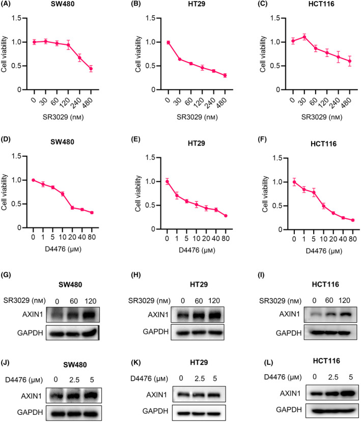
Casein kinase 1ε (CK1ε) and axis inhibitor 1 (AXIN1) are crucial components of the β-catenin destruction complex in canonical Wnt signaling. CK1ε has been shown to interact with AXIN1, but its physiological function and role in tumorigenesis remain unknown. In this study, we found that CK1δ/ε inhibitors significantly enhanced AXIN1 protein level in colorectal cancer (CRC) cells through targeting CK1ε. Mechanistically, CK1ε promoted AXIN1 degradation by the ubiquitin-proteasome pathway by promoting the interaction of E3 ubiquitin-protein ligase SIAH1 with AXIN1. Genetic or pharmacological inhibition of CK1ε and knockdown of SIAH1 downregulated the expression of Wnt/β-catenin-dependent genes, suppressed the viability of CRC cells, and restrained tumorigenesis and progression of CRC in vitro and in vivo. In summary, our results demonstrate that CK1ε exerted its oncogenic role in CRC occurrence and progression by regulating the stability of AXIN1. These findings reveal a novel mechanism by which CK1ε regulates the Wnt/β-catenin signaling pathway and highlight the therapeutic potential of targeting the CK1ε/SIAH1 axis in CRC.

Keywords: AXIN1; CK1ε; SIAH1; Wnt/β‐catenin; colorectal cancer; ubiquitination.


Conflict of interest statement

The authors declare no conflict of interest.

Figures

Fig. 1
Fig. 1
CK1δ/ε inhibitors induce AXIN1 protein stabilization in colorectal cancer cells. (A–C) The MTT assay was performed to assess the cell viability of SW480, HT29 and HCT116 cells exposed to different concentrations of SR3029 (0, 30, 60, 120, 240 and 480 nm) for 24 h (n = 5 independent experiments). (D–F) The MTT assay was performed to assess the viability of SW480, HT29 and HCT116 cells exposed to different concentrations of D4476 (0, 1, 5,10, 20, 40 and 80 μm) for 24 h (n = 5 independent experiments). (G–I) Immunoblot analysis of lysates of SW480, HT29 and HCT116 cells treated with 0, 60 and 120 nm SR3029 for 24 h before harvesting. (J–L) Immunoblot analysis of lysates of SW480, HT29 and HCT116 cells treated with 0, 2.5 and 5 μm D4476 for 24 h before harvesting. Shown is one representative of three independent experiments. Values are shown as means ± SD.
Fig. 2
Fig. 2
CK1ε regulates the protein expression of AXIN1. (A) Immunoblot analysis of lysates of HEK293T cells infected with shCtrl, shCK1ε‐1, shCK1ε‐2, shCK1δ‐1 and shCK1δ‐2 lentivirus. (B) Immunoblot analysis of lysates of HEK293T cells transfected with FLAG‐AXIN1 and gradient concentrations of GFP‐CK1ε (0, 0.1, 0.25, 0.5 and 1 μg) plasmid. (C) Immunoblot analysis of lysates of control cells and CK1ε‐deficient HEK293T cells (infected with lentivirus containing pBOBI‐CK1ε or pBOBI vector as a control) transfected with indicated plasmids encoding Flag‐tagged AXIN1. (D–F) Immunoblot analysis of lysates of SW480, HT29 and HCT116 cells infected with shCtrl, shCK1ε‐1, shCK1ε‐2, shCK1δ‐1 and shCK1δ‐2 lentivirus. Shown is one representative of three independent experiments.
Fig. 3
Fig. 3
CSNK1E knockdown reduces the viability and colon formation ability in CRC cells through attenuating Wnt/β‐catenin signaling. SW480 and HT29 cells were infected with the shCtrl or shCK1ε lentivirus respectively. (A, C) MTT assay was used to detect cell viability (n = 5 independent experiments). (B, D) Colony formation assay was performed to evaluate colony formation ability. Graphical representation of quantitative data showed the relative number of colonies formed (n = 3 independent experiments). (E, F) RT‐qPCR was performed to detect the mRNA expression of Wnt target genes (CYCLIND1, AXIN2, DKK1, and FIBRONECTIN). Quantification of mRNA level was normalized to GAPDH (n = 3 independent experiments). Values are shown as means ± SD. *P < 0.05, **P < 0.01, ***P < 0.001; Student's t test.
Fig. 4
Fig. 4
CK1ε regulates AXIN1 stability via the ubiquitin‐proteasome pathway. (A) Immunoblot analysis of lysates of HEK293T cells transfected with various indicated plasmids encoding FLAG‐AXIN1 and GFP‐CK1ε and then treated with 100 mg·mL−1 CHX for 0, 3, 6, and 9 h before harvesting. (B) Immunoblot analysis of lysates of HEK293T cells treated with or without 10 μm MG132 for 8 h, 50 nm CQ, 10 nm Baf‐A1 for 24 h with Wnt3a conditioned medium (Wnt3a‐CM) before harvesting. (C) Immunoblot analysis of lysates of HEK293T cells transfected with various indicated plasmids encoding FLAG‐AXIN1 and GFP‐CK1ε and then treated with 10 μm MG132 or DMSO for 8 h before harvesting. (D) Immunoprecipitation and immunoblot analysis of lysates of HEK293T cells transfected with various indicated plasmids encoding FLAG‐AXIN1, GFP‐CK1ε and HA‐Ub, and then treated with 10 μm MG132 for 8 h before harvesting. (E, F) Immunoprecipitation and immunoblot analysis of lysates of control or CSNK1E‐deficient cells treated with 10 μm MG132 for 8 h before harvesting. Shown is one representative of three independent experiments.
Fig. 5
Fig. 5
CK1ε promotes the interaction of SIAH1 with AXIN1. (A, B) Immunoprecipitation and immunoblot analyses of lysates of HEK293T cells transfected with various indicated plasmids encoding GFP‐CK1ε and HA‐SIAH1. (A) IP was performed with FLAG‐tagged beads. (B) IP was performed with GFP‐tagged beads. (C) Confocal microscopy of HEK293T cells transfected with plasmids encoding GFP‐CK1ε and HA‐SIAH1, scale bars = 15 μm. (D) Immunoprecipitation and immunoblot analyses of lysates of HEK293T cell were performed using control IgG or anti‐AXIN1 antibody. (E) Confocal microscopy of HEK293T cells transfected with plasmids encoding V5‐CK1ε, HA‐SIAH1 and GFP‐AXIN1, scale bars = 15 μm. (F) Immunoprecipitation and immunoblot analyses of lysates of HEK293T cells transfected with various indicated plasmids encoding GFP‐CK1ε, HA‐SIAH1 and FLAG‐AXIN1. (G) Immunoprecipitation and immunoblot analyses of lysates of CSNK1E‐deficient HEK293T cells or control cells transfected with indicated plasmids encoding HA‐SIAH1 and FLAG‐AXIN1. (H) Immunoprecipitation and immunoblot analyses of lysates of HEK293T cells transfected with various indicated plasmids encoding HA‐SIAH1 and FLAG‐AXIN1 and treated with DMSO or SR3029 (100 nm) 24 h. Shown is one representative of three independent experiments.
Fig. 6
Fig. 6
CK1ε synergizes with SIAH1 to promote AXIN1 ubiquitination and degradation. (A) Immunoblot analysis of lysates of HEK293T cells transfected with various indicated plasmids encoding HA‐SIAH1, GFP‐CK1ε and FLAG‐AXIN1. (B) Immunoblot analysis of lysates of CSNK1E‐deficient HEK293T cells or control cells transfected with indicated plasmids encoding HA‐SIAH1 and FLAG‐AXIN1. (C) Immunoblot analysis of lysates of CSNK1E‐deficient HEK293T cells or control cells infected with shCtrl or shSIAH1 lentivirus (n = 3 independent experiments). (D, E) Immunoblot analysis of lysates of SW480 and HCT116 cells infected with shCtrl or shSIAH1 lentivirus and then treated with 100 nm SR3029 for 24 h before harvesting. (F) Immunoprecipitation and immunoblot analyses of lysates of HEK293T cells transfected with various indicated plasmids encoding HA‐SIAH1, FLAG‐AXIN1 and GFP‐CK1ε and then treated with 10 μm MG132 for 8 h before harvesting. (G) The SuperTOPFlash reporter gene was transfected into HEK293T cells with SIAH1 or control plasmids together with gradient concentrations of CK1ε plasmid (n = 4 independent experiments). Shown is one representative of at least three independent experiments. Values are shown as means ± SD. *P < 0.05, **P < 0.01, ***P < 0.001; Student's t test.
Fig. 7
Fig. 7
SR3029 collaborates with knockdown of SIAH1 to inhibit cell viability and colony formation ability via downregulating Wnt/β‐catenin signaling in CRC cells. SW480 and HCT116 cells were infected with the shCtrl or shSIAH1 lentivirus, then treated with DMSO or SR3029 (100 nm) for 24 h (A, B, E, F), 48 h (A, B) or 12 days (C, D), respectively. (A, B) MTT assay was used to detect cell viability (n = 5 independent experiments). (C, D) Colony formation assay was performed to evaluate the cell proliferation. Graphical representation of quantitative data showed the relative number of colonies formed (n = 3 independent experiments). (E, F) RT‐qPCR was performed to detect mRNA expression of Wnt target genes (CYCLIND1, AXIN2, TWIST). Quantification of mRNA level was normalized to GAPDH (n = 3 independent experiments). Values are shown as means ± SD. *P < 0.05, **P < 0.01, ***P < 0.001; Student's t test.
Fig. 8
Fig. 8
SR3029 enhances the suppressive effect of Siah1α knockdown on colorectal tumor growth in vivo by stabilizing AXIN1. (A) CRC xenograft model. Siah1α‐deficient MC38 cells and parental counterparts were subcutaneously (s.c.) implanted into the right back of 8‐week‐old male C57BL/6J mice to generate a xenograft tumor model. When the tumors reached about 50 mm3, mice were randomly divided into two groups and intraperitoneally (i.p.) injected with the vehicle (10% DMSO/40% PEG‐300/5% Tween‐80/45% saline) or SR3029 (10 mg·kg−1) every 3 days. Tumor sizes were measured with a caliper and tumor volumes were calculated using the formula: 0.5 × length × width2. At 9 days after SR3029 treatment, mice were sacrificed, and tumors were collected and photographed. (B) Images of tumors from each experimental group. (C) Mean tumor volume in each experimental group (n = 5 independent experiments). (D) Mean tumor weight in each experimental group (n = 5 independent experiments). (E) Immunoblot analysis of AXIN1 in each experimental group. (F) RT‐qPCR analysis of Wnt target genes (CyclinD1, Axin2) in each experimental group (n = 3 independent experiments). (G) H&E, AXIN1 and CYCLIND1 staining of each experimental group, scale bars = 50 μm. Shown is one representative of three independent experiments. Values are shown as means ± SEM. *P < 0.05, **P < 0.01, ***P < 0.001; Two‐way ANOVA for (C); Student's t test for (D) and (F).
Fig. 9
Fig. 9
Molecular model depicting the CK1ε‐SIAH1‐AXIN1 regulatory module in CRC cells. Schematic representation of the role of CK1ε in the regulation of AXIN1 degradation via the ubiquitin‐proteasome pathway. CK1ε promoted AXIN1 degradation by the ubiquitin‐proteasome pathway via promoting the interaction of E3 ubiquitin ligase SIAH1 with AXIN1. CK1δ/ε inhibitors could significantly enhance AXIN1 protein level through targeting CK1ε.

Similar articles

Cited by

References

    1. Sung H, Ferlay J, Siegel RL, Laversanne M, Soerjomataram I, Jemal A, et al. Global cancer statistics 2020: GLOBOCAN estimates of incidence and mortality worldwide for 36 cancers in 185 countries. CA Cancer J Clin. 2021;71(3):209–249. - PubMed
    1. Cho YH, Ro EJ, Yoon JS, Mizutani T, Kang DW, Park JC, et al. 5‐FU promotes stemness of colorectal cancer via p53‐mediated WNT/beta‐catenin pathway activation. Nat Commun. 2020;11(1):5321. - PMC - PubMed
    1. Liu Z, Ma T, Duan J, Liu X, Liu L. MicroRNA‐223‐induced inhibition of the FBXW7 gene affects the proliferation and apoptosis of colorectal cancer cells via the notch and Akt/mTOR pathways. Mol Med Rep. 2021;23(2):154. - PMC - PubMed
    1. Schloss J, Ryan K, Steel A. Corrigendum to a randomised, double‐blind, placebo‐controlled clinical trial found that a novel herbal formula UROX(R) BEDTIME BUDDY assisted children for the treatment of nocturnal enuresis. Phytomedicine. 2022;99:153992. - PubMed
    1. Javed Z, Javed Iqbal M, Rasheed A, Sadia H, Raza S, Irshad A, et al. Regulation of hedgehog signaling by miRNAs and nanoformulations: a possible therapeutic solution for colorectal cancer. Front Oncol. 2020;10:607607. - PMC - PubMed

MeSH terms