

A Cone Bean Computer Tomography Investigation of the Newly Formed Mandibular Anterior Ridge following the Treatment of an Extended Comminuted Fracture: A 12-Year Follow-Up
- PMID: 38419613
- PMCID: PMC10901576
- DOI: 10.1155/2024/1824016
A Cone Bean Computer Tomography Investigation of the Newly Formed Mandibular Anterior Ridge following the Treatment of an Extended Comminuted Fracture: A 12-Year Follow-Up
Abstract
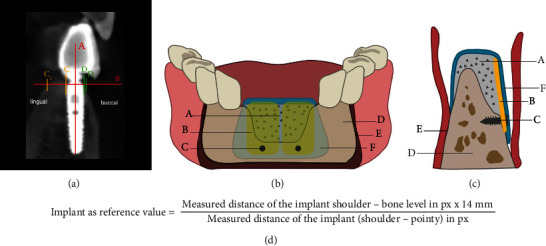
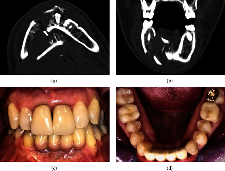
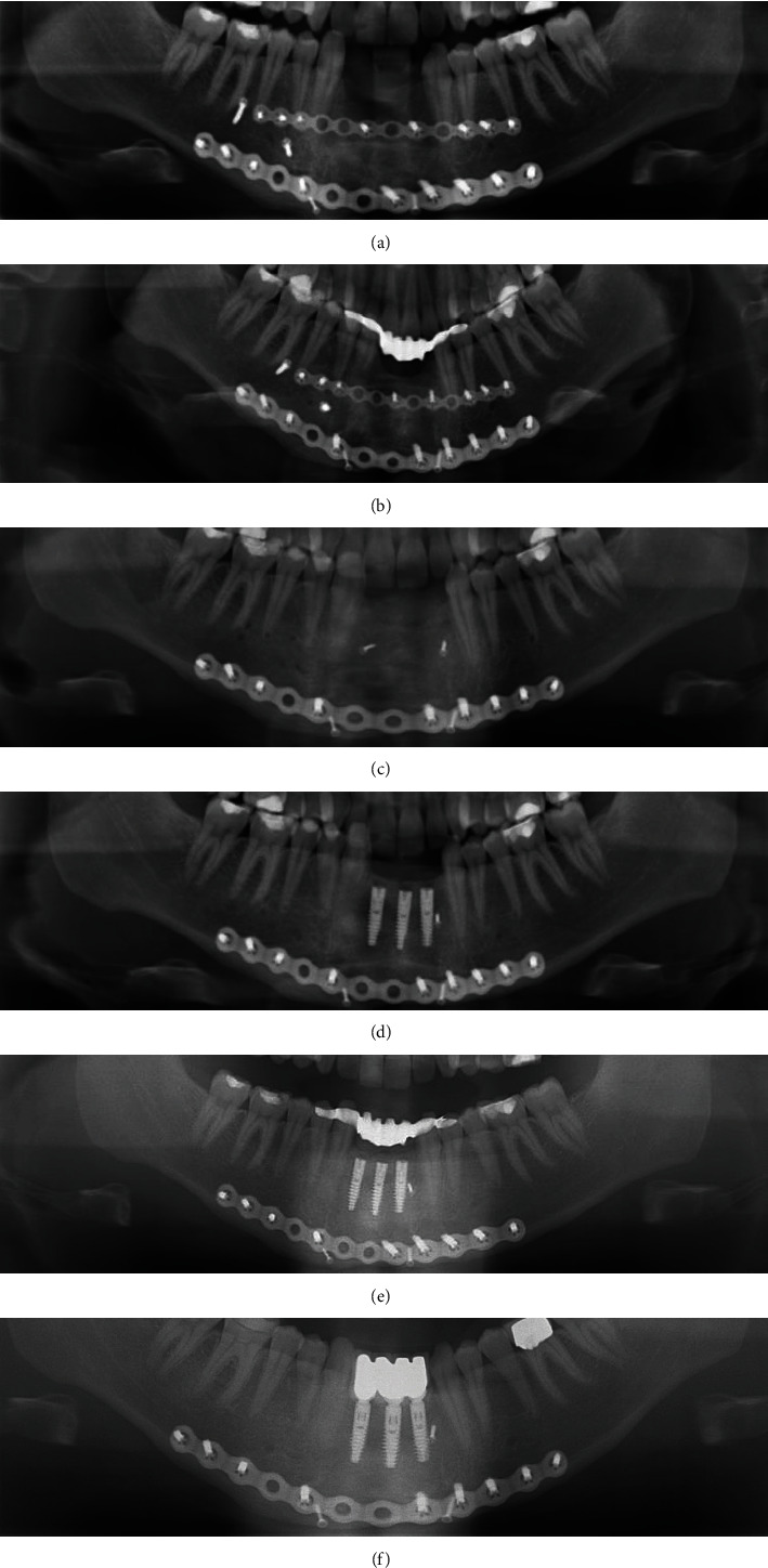
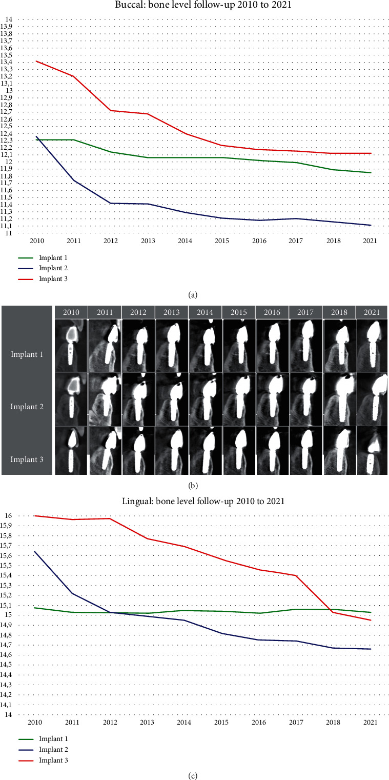
Introduction: Extensive comminuted fractures are associated with tooth loss that ultimately leads to dimensional changes in the hard and soft tissues of the alveolar ridge. Reconstruction of the lost mandibular anterior ridge is very complex due to the natural curvature of the region. Case Presentation. In this case report, the combination of the modified shell technique with autologous bone plates and the guided bone regeneration (GBR) technique was performed on an 18-year-old patient after a comminuted fracture, to ensure new bone formation in the anterior ridge with a natural curvature. After the treatment progressed without complications, three dental implants were placed. Annual cone beam computed tomography (CBCT) images were obtained and evaluated using the GNU Image Manipulation Program (GIMP© 2.10). This allowed measurements of the buccal and lingual bone around the implants, showing the annual bone loss in a twelve-year observation period. Discussion. The treatment of the comminuted fracture and the combination of the modified shell technique with autologous bone plates, the GBR technique, and implant placement can be considered successful. The three dental implants were osseointegrated in 2010, with the buccal bone level averaging 1.31 mm below the implant shoulder and the lingual bone level 1.57 mm above the implant shoulder. In 2021, the measurements showed a bone loss of 0.99 mm at the buccal implant shoulder and 0.69 mm at the lingual implant shoulder.
Conclusion: The combination of the modified shell technique with autologous bone plates and the GBR technique is a reliable method to ensure new bone formation in the anterior ridge. The use of CBCT is an excellent method to evaluate bone resorption around dental implants, but due to minimal bone resorption in the observation period, an annual CBCT examination is exaggerated.
Copyright © 2024 Pascal Grün et al.
Conflict of interest statement
The authors declare that they have no conflicts of interest.
Figures
Similar articles
-
Buccal bone thickness adjacent to virtual dental implants following guided bone regeneration.J Periodontol. 2019 Jun;90(6):595-607. doi: 10.1002/JPER.18-0304. Epub 2019 Jan 10. J Periodontol. 2019. PMID: 30578550 Clinical Trial.
-
Ridge expansion alone or in combination with guided bone regeneration to facilitate implant placement in narrow alveolar ridges: a retrospective study.Clin Oral Implants Res. 2015 Feb;26(2):204-11. doi: 10.1111/clr.12317. Epub 2013 Dec 16. Clin Oral Implants Res. 2015. PMID: 24330035
-
Evaluation of post-implant buccal bone resorption using cone beam computed tomography: a clinical pilot study.Int J Oral Maxillofac Implants. 2012 Sep-Oct;27(5):1249-57. Int J Oral Maxillofac Implants. 2012. PMID: 23057042
-
Ridge preservation after ridge expansion with simultaneous guided bone regeneration: a preclinical study.Clin Oral Implants Res. 2016 Nov;27(11):e116-e124. doi: 10.1111/clr.12574. Epub 2015 Mar 9. Clin Oral Implants Res. 2016. PMID: 25754343
-
Buccal bone crest dynamics after immediate implant placement and ridge preservation techniques: review of morphometric studies in animals.Implant Dent. 2013 Apr;22(2):155-60. doi: 10.1097/ID.0b013e318287a947. Implant Dent. 2013. PMID: 23462592 Review.
References
Publication types
LinkOut - more resources
Full Text Sources
