

Metabolic dynamics in astrocytes and microglia during post-natal development and their implications for autism spectrum disorders
- PMID: 38419654
- PMCID: PMC10899402
- DOI: 10.3389/fncel.2024.1354259
Metabolic dynamics in astrocytes and microglia during post-natal development and their implications for autism spectrum disorders
Abstract
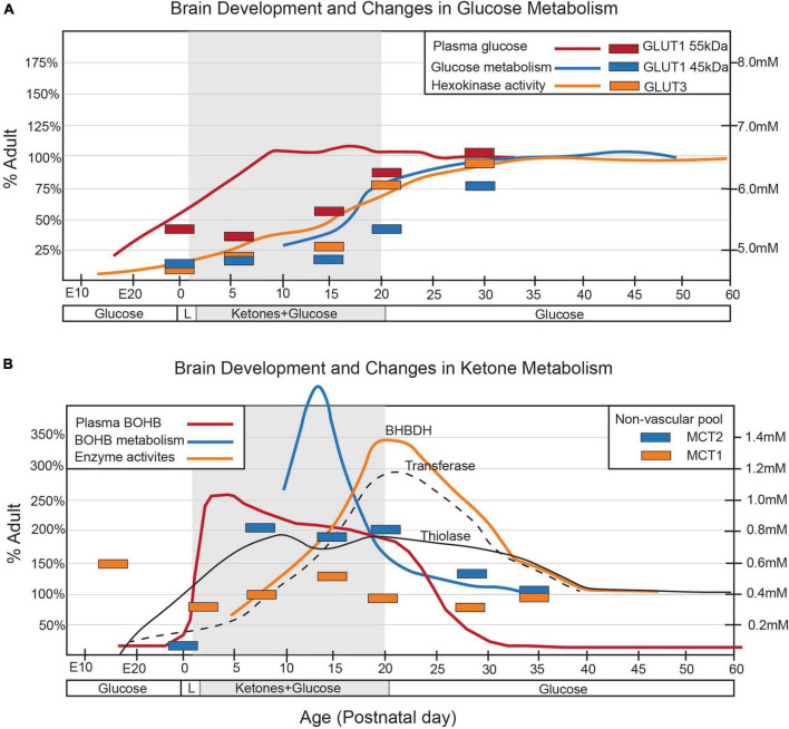
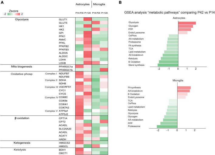
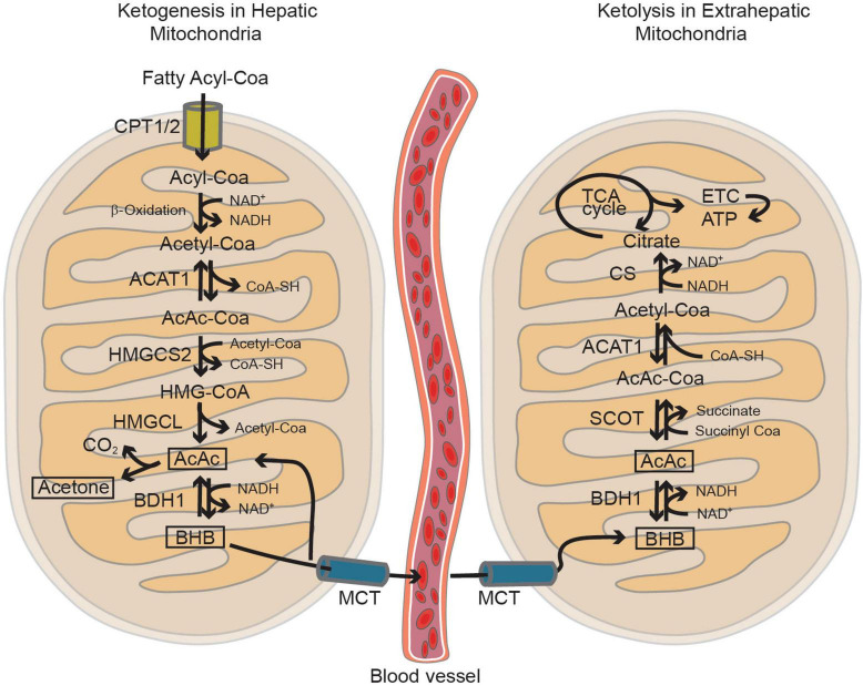
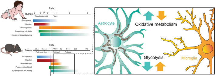
Autism Spectrum Disorder (ASD) is a complex neurodevelopmental condition characterized by elusive underlying mechanisms. Recent attention has focused on the involvement of astrocytes and microglia in ASD pathology. These glial cells play pivotal roles in maintaining neuronal homeostasis, including the regulation of metabolism. Emerging evidence suggests a potential association between ASD and inborn errors of metabolism. Therefore, gaining a comprehensive understanding of the functions of microglia and astrocytes in ASD is crucial for the development of effective therapeutic interventions. This review aims to provide a summary of the metabolism of astrocytes and microglia during post-natal development and the evidence of disrupted metabolic pathways in ASD, with particular emphasis on those potentially important for the regulation of neuronal post-natal maturation by astrocytes and microglia.
Keywords: astrocyte; autism spectrum disorders; brain metabolism; fatty acid oxidation; lactate; lipids; microglia; mitochondria.
Copyright © 2024 Cantando, Centofanti, D’Alessandro, Limatola and Bezzi.
Conflict of interest statement
The authors declare that the research was conducted in the absence of any commercial or financial relationships that could be construed as a potential conflict of interest.
Figures
Similar articles
-
Microglia and astrocytes underlie neuroinflammation and synaptic susceptibility in autism spectrum disorder.Front Neurosci. 2023 Mar 20;17:1125428. doi: 10.3389/fnins.2023.1125428. eCollection 2023. Front Neurosci. 2023. PMID: 37021129 Free PMC article. Review.
-
Pathology and Astrocytes in Autism.Neuropsychiatr Dis Treat. 2023 Apr 12;19:841-850. doi: 10.2147/NDT.S390053. eCollection 2023. Neuropsychiatr Dis Treat. 2023. PMID: 37077706 Free PMC article. Review.
-
Microglia at the Tripartite Synapse during Postnatal Development: Implications for Autism Spectrum Disorders and Schizophrenia.Cells. 2023 Dec 13;12(24):2827. doi: 10.3390/cells12242827. Cells. 2023. PMID: 38132147 Free PMC article. Review.
-
Microglia Modulate Neurodevelopment in Autism Spectrum Disorder and Schizophrenia.Int J Mol Sci. 2023 Dec 9;24(24):17297. doi: 10.3390/ijms242417297. Int J Mol Sci. 2023. PMID: 38139124 Free PMC article. Review.
-
The Role of Glial Cells in Autism Spectrum Disorder: Molecular Mechanisms and Therapeutic Approaches.CNS Neurol Disord Drug Targets. 2025 Jan 3. doi: 10.2174/0118715273337007241115102118. Online ahead of print. CNS Neurol Disord Drug Targets. 2025. PMID: 39773050
Cited by
-
Dual lineage origins contribute to neocortical astrocyte diversity.Nat Commun. 2025 Jul 30;16(1):6992. doi: 10.1038/s41467-025-61829-4. Nat Commun. 2025. PMID: 40738880 Free PMC article.
-
Metabolic Reprogramming of Astrocytes in Pathological Conditions: Implications for Neurodegenerative Diseases.Int J Mol Sci. 2024 Aug 16;25(16):8922. doi: 10.3390/ijms25168922. Int J Mol Sci. 2024. PMID: 39201607 Free PMC article. Review.
-
Lactate reprograms glioblastoma immunity through CBX3-regulated histone lactylation.J Clin Invest. 2024 Nov 15;134(22):e176851. doi: 10.1172/JCI176851. J Clin Invest. 2024. PMID: 39545414 Free PMC article.
-
Astrocyte Dysfunctions in Obsessive Compulsive Disorder: Rethinking Neurobiology and Therapeutic Targets.J Neurochem. 2025 May;169(5):e70092. doi: 10.1111/jnc.70092. J Neurochem. 2025. PMID: 40400176 Free PMC article. Review.
-
Correlation of biochemical markers and inflammatory cytokines in autism spectrum disorder (ASD).BMC Pediatr. 2024 Nov 1;24(1):696. doi: 10.1186/s12887-024-05182-3. BMC Pediatr. 2024. PMID: 39487445 Free PMC article.
References
Publication types
LinkOut - more resources
Full Text Sources