

Damage-evoked signals in cochlear neurons and supporting cells
- PMID: 38419694
- PMCID: PMC10899329
- DOI: 10.3389/fneur.2024.1361747
Damage-evoked signals in cochlear neurons and supporting cells
Abstract
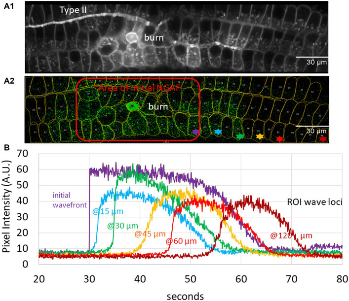
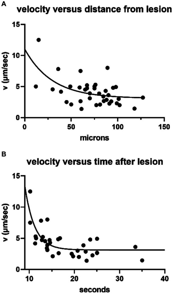
In addition to hearing loss, damage to the cochlea can lead to gain of function pathologies such as hyperacusis. It has been proposed that painful hyperacusis, noxacusis, may be carried to the central nervous system by type II cochlear afferents, sparse, unmyelinated neurons that share morphological and neurochemical traits with nociceptive C-fibers of the somatic nervous system. Also like in skin, damage elicits spreading calcium waves within cochlear epithelia. These are mediated by extracellular ATP combined with IP3-driven release from intracellular calcium stores. Type II afferents are excited by ATP released from damaged epithelia. Thus, the genesis and propagation of epithelial calcium waves is central to cochlear pathology, and presumably hyperacusis. Damage-evoked signals in type II afferents and epithelial cells have been recorded in cochlear explants or semi-intact otic capsules. These efforts have included intracellular electrical recording, use of fluorescent calcium indicators, and visualization of an activity-dependent, intrinsic fluorescent signal. Of relevance to hyperacusis, prior noise-induced hearing loss leads to the generation of prolonged and repetitive activity in type II neurons and surrounding epithelia.
Keywords: calcium waves; cochlea; epithelia; hyperacusis; trauma; type II afferent.
Copyright © 2024 Wood, Nowak and Fuchs.
Conflict of interest statement
The authors declare that the research was conducted in the absence of any commercial or financial relationships that could be construed as a potential conflict of interest.
Figures
Similar articles
-
Unmyelinated type II afferent neurons report cochlear damage.Proc Natl Acad Sci U S A. 2015 Nov 24;112(47):14723-7. doi: 10.1073/pnas.1515228112. Epub 2015 Nov 9. Proc Natl Acad Sci U S A. 2015. PMID: 26553995 Free PMC article.
-
Prior Acoustic Trauma Alters Type II Afferent Activity in the Mouse Cochlea.eNeuro. 2021 Nov 11;8(6):ENEURO.0383-21.2021. doi: 10.1523/ENEURO.0383-21.2021. Print 2021 Nov-Dec. eNeuro. 2021. PMID: 34607806 Free PMC article.
-
Opposing expression gradients of calcitonin-related polypeptide alpha (Calca/Cgrpα) and tyrosine hydroxylase (Th) in type II afferent neurons of the mouse cochlea.J Comp Neurol. 2018 Feb 15;526(3):425-438. doi: 10.1002/cne.24341. Epub 2017 Nov 13. J Comp Neurol. 2018. PMID: 29055051 Free PMC article.
-
Advances in the neurobiology of hearing disorders: recent developments regarding the basis of tinnitus and hyperacusis.Prog Neurobiol. 2013 Dec;111:17-33. doi: 10.1016/j.pneurobio.2013.08.002. Epub 2013 Sep 6. Prog Neurobiol. 2013. PMID: 24012803 Review.
-
Inner Hair Cell Loss Disrupts Hearing and Cochlear Function Leading to Sensory Deprivation and Enhanced Central Auditory Gain.Front Neurosci. 2017 Jan 18;10:621. doi: 10.3389/fnins.2016.00621. eCollection 2016. Front Neurosci. 2017. PMID: 28149271 Free PMC article. Review.
Cited by
-
Clinical phenotype and management of sound-induced pain: Insights from adults with pain hyperacusis.J Pain. 2025 Feb;27:104741. doi: 10.1016/j.jpain.2024.104741. Epub 2024 Nov 23. J Pain. 2025. PMID: 39586560
-
Virally mediated enhancement of efferent inhibition reduces acoustic trauma in wild-type murine cochleas.Mol Ther Methods Clin Dev. 2025 Mar 21;33(2):101455. doi: 10.1016/j.omtm.2025.101455. eCollection 2025 Jun 12. Mol Ther Methods Clin Dev. 2025. PMID: 40236498 Free PMC article.
-
Virally-Mediated Enhancement of Efferent Inhibition Reduces Acoustic Trauma in Wild Type Murine Cochleas.bioRxiv [Preprint]. 2024 Sep 15:2024.09.12.612688. doi: 10.1101/2024.09.12.612688. bioRxiv. 2024. Update in: Mol Ther Methods Clin Dev. 2025 Mar 21;33(2):101455. doi: 10.1016/j.omtm.2025.101455. PMID: 39314296 Free PMC article. Updated. Preprint.
-
Clinical phenotype and management of sound-induced pain: Insights from adults with pain hyperacusis.medRxiv [Preprint]. 2024 Jun 20:2024.06.19.24309185. doi: 10.1101/2024.06.19.24309185. medRxiv. 2024. Update in: J Pain. 2025 Feb;27:104741. doi: 10.1016/j.jpain.2024.104741. PMID: 38946957 Free PMC article. Updated. Preprint.
References
-
- Weisz CJC, Williams SG, Eckard CS, Divito CB, Ferreira DW, Fantetti KN, et al. . Outer hair cell glutamate signaling through type II spiral ganglion afferents activates neurons in the Cochlear nucleus in response to nondamaging sounds. J Neurosci. (2021) 41:2930–43. doi: 10.1523/JNEUROSCI.0619-20.2021, PMID: - DOI - PMC - PubMed
Publication types
Grants and funding
LinkOut - more resources
Full Text Sources