

The imaging dynamic changes in the malignant transformation of an epidermoid cyst: a case report and literature review
- PMID: 38419698
- PMCID: PMC10900506
- DOI: 10.3389/fneur.2024.1349044
The imaging dynamic changes in the malignant transformation of an epidermoid cyst: a case report and literature review
Abstract
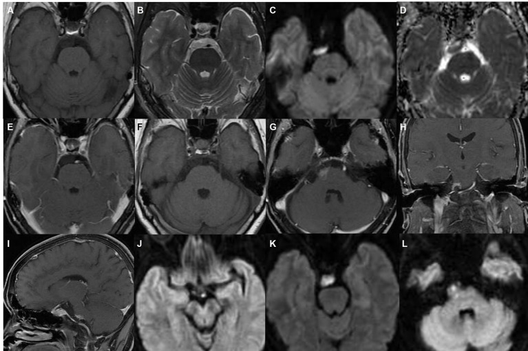
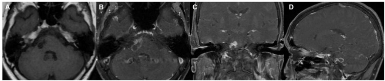
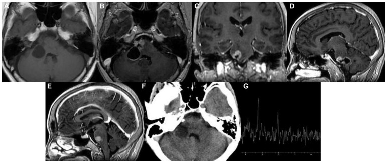
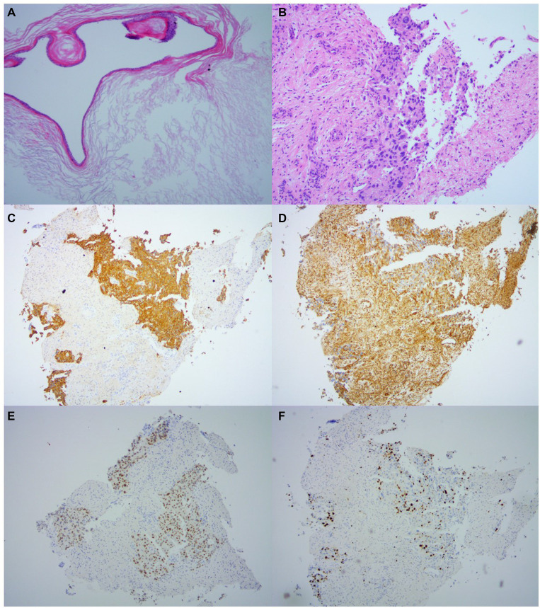
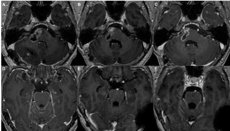
Malignant transformation of epidermoid cysts is a rare complication. Most of the previously reported cases have involved postoperative malignant transformations. We present a case of malignant transformation of a nonpostoperative epidermoid tumor into squamous cell carcinoma (SCC) that occurred in a 61-year-old Chinese woman. The patient's initial cranial MRI scan showed an epidermoid cyst with marginal enhancement in the pre-pontine cistern, and the lesion gradually enlarged after 10 months. A craniotomy was performed using to remove part of the tumor via the right retrosigmoid approach, and postoperative pathology confirmed that the transformation of the epidermoid cyst was malignant. Our case study suggests that the possibility of malignant transformation of epidermoid cyst should not be ignored on the basis of enhanced imaging features, regardless of whether they are nodular, annular, or patchy, as is the case for inflammation. Strict follow-up is required for early detection of malignant transformation to prompt correspondingly early clinical treatment.
Keywords: epidermoid cysts; follow-up; malignant transformation; pre-pontine cistern; squamous cell carcinoma.
Copyright © 2024 Yang, Hu, Li, Xu, Zhang, Huang, Yang and Zhang.
Conflict of interest statement
The authors declare that the research was conducted in the absence of any commercial or financial relationships that could be construed as a potential conflict of interest.
Figures
Similar articles
-
Cerebellar Squamous Cell Carcinoma Due to Malignant Transformation of Cerebellopontine Angle Epidermoid Cyst, Report an Interesting Case and Review the Literature.Prague Med Rep. 2019;120(2-3):95-102. doi: 10.14712/23362936.2019.14. Prague Med Rep. 2019. PMID: 31586508 Review.
-
Radio-pathological characteristics of malignant transformation of an epidermoid cyst in the cerebellopontine angle: A case report.Surg Neurol Int. 2022 Apr 8;13:135. doi: 10.25259/SNI_1226_2021. eCollection 2022. Surg Neurol Int. 2022. PMID: 35509542 Free PMC article.
-
Malignant transformation of an epidermoid cyst in the cerebellopontine angle.J Korean Neurosurg Soc. 2012 Aug;52(2):148-51. doi: 10.3340/jkns.2012.52.2.148. Epub 2012 Aug 31. J Korean Neurosurg Soc. 2012. PMID: 23091675 Free PMC article.
-
Malignant transformation 20 years after partial removal of intracranial epidermoid cyst--case report.Neurol Med Chir (Tokyo). 2010;50(3):236-9. doi: 10.2176/nmc.50.236. Neurol Med Chir (Tokyo). 2010. PMID: 20339276
-
A Rare Transformation of Epidermoid Cyst into Squamous Cell Carcinoma: A Case Report with Literature Review.Am J Case Rep. 2019 Aug 3;20:1141-1143. doi: 10.12659/AJCR.912828. Am J Case Rep. 2019. PMID: 31375657 Free PMC article. Review.
References
-
- Mliyh L, Di Perri D, Onofrj V. Intracranial squamous cell carcinoma of the cerebello-pontine angle mimicking a cystic acoustic schwannoma. A case report with discussion of differential diagnosis and review of literature. Radiol Case Rep. (2023) 18:753–6. doi: 10.1016/j.radcr.2022.11.048, PMID: - DOI - PMC - PubMed
Publication types
LinkOut - more resources
Full Text Sources
Research Materials