. 2024 Jan 30;6(5):1497-1506.
doi: 10.1039/d3na00751k. eCollection 2024 Feb 27.

Nanogold-coated stent facilitated non-invasive photothermal ablation of stent thrombosis and restoration of blood flow

Affiliations

Nanogold-coated stent facilitated non-invasive photothermal ablation of stent thrombosis and restoration of blood flow

Nitesh Singh et al. Nanoscale Adv. .

Abstract

In-stent restenosis (ISR) and stent thrombosis (ST) are the most serious complications of coronary angioplasty and stenting. Although the evolution of drug-eluting stents (DES) has significantly restricted the incidence of ISR, they are associated with an enhanced risk of ST. In the present study, we explore the photothermal ablation of a thrombus using a nano-enhanced thermogenic stent (NETS) as a modality for revascularization following ST. The photothermal activity of NETS, fabricated by coating bare metal stents with gold nanorods generating a thin plasmonic film of gold, was found to be effective in rarefying clots formed within the stent lumen in various in vitro assays including those under conditions mimicking blood flow. NETS implanted in the rat common carotid artery generated heat following exposure to a NIR-laser that led to effective restoration of blood flow within the occluded vessel in a model of ferric chloride-induced thrombosis. Our results present a proof-of-concept for a novel photothermal ablation approach by employing coated stents in the non-invasive management of ST.


Conflict of interest statement

The authors declare no competing interests.

Figures

Fig. 1
Fig. 1. Scanning electron micrographs of surface morphologies of (A) bare (uncoated) and (B) coated stents. Magnifications: upper panels, 25× (scale, 200 μm); lower panels, 400× (scale, 1 μm).
Fig. 2
Fig. 2. EDX analyses of (A) bare (uncoated) and (B) coated stents.
Fig. 3
Fig. 3. Photothermal ablation of in-stent/peri-stent clots exposed to the NIR laser for 30 min. Clots were generated by addition of 2–5 mM CaCl2 and 1 U ml−1 thrombin to a solution of fibrinogen (1 mg ml−1) carrying the thermogenic stents, followed by irradiation with the NIR (808 nm) laser at a power density 1.05 W cm−2. Whenever required, the fluorescent fibrin clot was generated by addition of Alexa Fluor 488 – conjugated fibrinogen (10% v/v) to the above solution. Drabkin's assay (A) and (B) and methylene blue assay (C) and (D) were carried out in order to evaluate the extent of clot lysis. (E) In-stent or peri-stent clot lysis analyzed employing an Alexa Fluor 488-labeled thrombus. (F) Lysis of a fluorescently labeled stent thrombus exposed to arterial hydrodynamic shear (1500 s−1). Data are representative of five different sets of experiments (mean ± SEM). *P < 0.05; **P < 0.01; ****P < 0.0001, vs. control.
Fig. 4
Fig. 4. FRAP analysis of a laser irradiated in-stent thrombus. (A–C) Confocal images of an in-stent fluorescent clot (10×) representing fluorescent (A), differential interference contrast (DIC) (B) and merged (C) images. (D–I) Pre-bleached, post-bleached and fluorescence recovery images (63×) of control and NIR laser-irradiated samples, as stated. Arrows indicate the regions of interest (ROI) on the fluorescent thrombus. (J) Kinetics of fluorescence recovery in control and NIR laser-treated samples.
Fig. 5
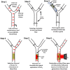
Fig. 5. Scheme for grafting a thermogenic stent in the common carotid artery of a rat, induction of a thrombus and photothermal thrombolysis.
Fig. 6
Fig. 6. Photothermal lysis of an in-stent thrombus and restoration of the blood flow. (A) Photograph of NETS implanted in the rat common carotid artery; (B) and (C) photoacoustic imaging of implanted NETS in B-mode (ultrasound) and PA-mode (photoacoustic), respectively. The arrow indicates the position of the stent. (D) Measurement of blood flow velocity in the rat common carotid artery with the implanted stent with a flow meter showing occlusion of blood flow due to the growing thrombus and subsequent restoration of flow following NIR laser irradiation. (E) Scatter dot plots representing the corresponding flow velocities after 15–20 min irradiation with the NIR laser or controls without laser exposure (n = 7 in each group). Each dot in the scatter plots represents an independent observation. **P < 0.01 vs. control. (F) and (G) Light microscopy of hematoxylin- and eosin-stained transverse sections of stented carotid segments harvested post-surgery from sacrificed animals with or without irradiation with the NIR laser (the arrow indicates reduction in thrombus size; scale, 100 μm).

Similar articles

References

    1. WHO, Cardiovascular diseases, https://www.who.int/health-topics/cardiovascular-diseases#tab=tab_1
    1. CDC, Heart Disease Deaths Vary by Sex, Race, and Ethnicity, https://www.cdc.gov/heartdisease/facts.htm#:∼:text=HeartDiseaseintheUnit...
    1. Simard T. Hibbert B. Ramirez F. D. Froeschl M. Chen Y. X. O'Brien E. R. Can. J. Cardiol. 2014;30:35–45. doi: 10.1016/j.cjca.2013.09.012. - DOI - PubMed
    1. Claessen B. E. Henriques J. P. Jaffer F. A. Mehran R. Piek J. J. Dangas G. D. JACC Cardiovasc. Interv. 2014;7:1081–1092. doi: 10.1016/j.jcin.2014.05.016. - DOI - PubMed
    1. Serruys P. W. Strauss B. H. Beatt K. J. Bertrand M. E. Puel J. Rickards A. F. Meier B. Goy J. J. Vogt P. Kappenberger L. et al. . N. Engl. J. Med. 1991;324:13–17. doi: 10.1056/NEJM199101033240103. - DOI - PubMed