

Radiomics for differentiation of gliomas from primary central nervous system lymphomas: a systematic review and meta-analysis
- PMID: 38420015
- PMCID: PMC10899458
- DOI: 10.3389/fonc.2024.1291861
Radiomics for differentiation of gliomas from primary central nervous system lymphomas: a systematic review and meta-analysis
Abstract
Background and objective: Numerous radiomics-based models have been proposed to discriminate between central nervous system (CNS) gliomas and primary central nervous system lymphomas (PCNSLs). Given the heterogeneity of the existing models, we aimed to define their overall performance and identify the most critical variables to pilot future algorithms.
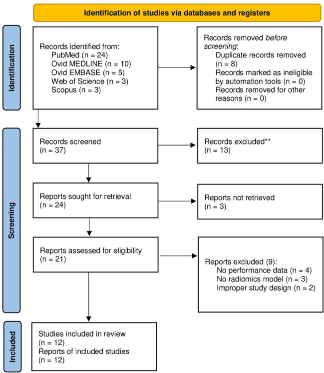
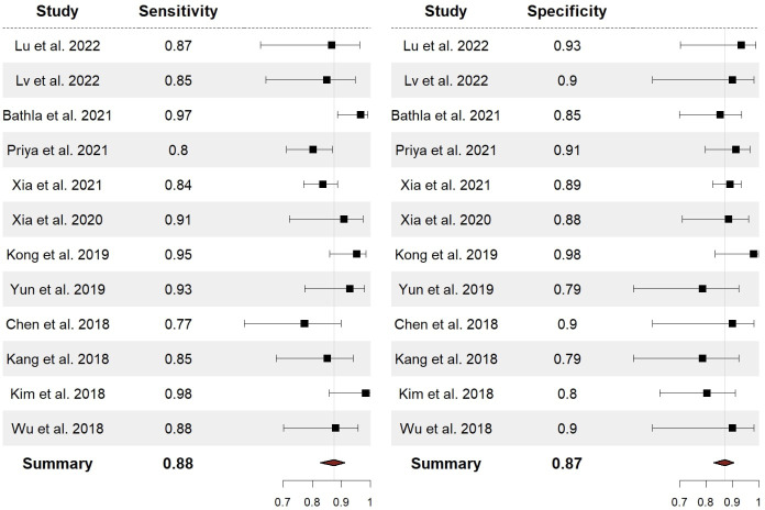
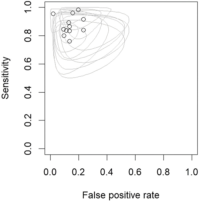
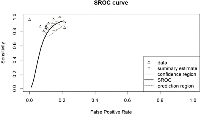
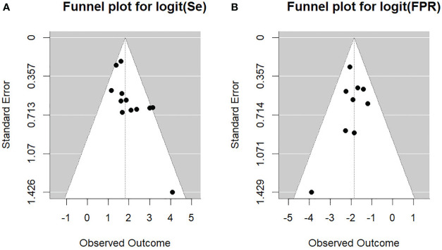
Methods: A systematic review of the literature and a meta-analysis were conducted, encompassing 12 studies and a total of 1779 patients, focusing on radiomics to differentiate gliomas from PCNSLs. A comprehensive literature search was performed through PubMed, Ovid MEDLINE, Ovid EMBASE, Web of Science, and Scopus databases. Overall sensitivity (SEN) and specificity (SPE) were estimated. Event rates were pooled using a random-effects meta-analysis, and the heterogeneity was assessed using the χ2 test.
Results: The overall SEN and SPE for differentiation between CNS gliomas and PCNSLs were 88% (95% CI = 0.83 - 0.91) and 87% (95% CI = 0.83 - 0.91), respectively. The best-performing features were the ones extracted from the Gray Level Run Length Matrix (GLRLM; ACC 97%), followed by those obtained from the Neighboring Gray Tone Difference Matrix (NGTDM; ACC 93%), and shape-based features (ACC 91%). The 18F-FDG-PET/CT was the best-performing imaging modality (ACC 97%), followed by the MRI CE-T1W (ACC 87% - 95%). Most studies applied a cross-validation analysis (92%).
Conclusion: The current SEN and SPE of radiomics to discriminate CNS gliomas from PCNSLs are high, making radiomics a helpful method to differentiate these tumor types. The best-performing features are the GLRLM, NGTDM, and shape-based features. The 18F-FDG-PET/CT imaging modality is the best-performing, while the MRI CE-T1W is the most used.
Keywords: gliomas; meta-analysis; primary central nervous system lymphomas; radiomics; systematic review.
Copyright © 2024 Garaba, Aslam, Ponzio, Panciani, Brinjikji, Fontanella and De Maria.
Conflict of interest statement
The authors declare that the research was conducted in the absence of any commercial or financial relationships that could be construed as a potential conflict of interest. The author(s) declared that they were an editorial board member of Frontiers, at the time of submission. This had no impact on the peer review process and the final decision.
Figures
Similar articles
-
Radiomics for Differentiation of Pediatric Posterior Fossa Tumors: A Meta-Analysis and Systematic Review of the Literature.Cancers (Basel). 2023 Dec 18;15(24):5891. doi: 10.3390/cancers15245891. Cancers (Basel). 2023. PMID: 38136435 Free PMC article. Review.
-
The Current Diagnostic Performance of MRI-Based Radiomics for Glioma Grading: A Meta-Analysis.J Integr Neurosci. 2024 May 14;23(5):100. doi: 10.31083/j.jin2305100. J Integr Neurosci. 2024. PMID: 38812383
-
Fully automated MR-based virtual biopsy of primary CNS lymphomas.Neurooncol Adv. 2024 Mar 14;6(1):vdae022. doi: 10.1093/noajnl/vdae022. eCollection 2024 Jan-Dec. Neurooncol Adv. 2024. PMID: 38516329 Free PMC article.
-
Use of 18F-FDG-PET/CT in differential diagnosis of primary central nervous system lymphoma and high-grade gliomas: A meta-analysis.Front Neurol. 2022 Aug 17;13:935459. doi: 10.3389/fneur.2022.935459. eCollection 2022. Front Neurol. 2022. PMID: 36061992 Free PMC article.
-
An integrative non-invasive malignant brain tumors classification and Ki-67 labeling index prediction pipeline with radiomics approach.Eur J Radiol. 2023 Jan;158:110639. doi: 10.1016/j.ejrad.2022.110639. Epub 2022 Nov 28. Eur J Radiol. 2023. PMID: 36463703
Cited by
-
Artificial Intelligence and Machine Learning in Neuroregeneration: A Systematic Review.Cureus. 2024 May 30;16(5):e61400. doi: 10.7759/cureus.61400. eCollection 2024 May. Cureus. 2024. PMID: 38953082 Free PMC article. Review.
References
Publication types
LinkOut - more resources
Full Text Sources