

Magnetic Resonance Imaging Features of Congenital Infantile Fibrosarcoma
- PMID: 38420085
- PMCID: PMC10899809
- DOI: 10.7759/cureus.53132
Magnetic Resonance Imaging Features of Congenital Infantile Fibrosarcoma
Abstract
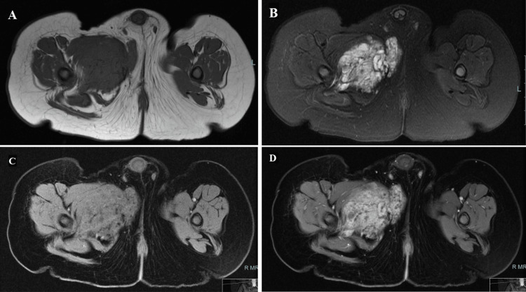
Background Congenital/infantile fibrosarcoma is a rare soft tissue tumor presented in early age of life. It should be considered in the differential diagnosis of the large soft tissue masses especially in the extremities at the age of infancy. These tumors frequently are misdiagnosed at birth as hemangioma. Histologically, they can resemble their adult counterparts and they are characterized by the chromosomal translocation t(12;15) (p13;q25) resulting in the ETV6-NTRK3 gene fusion. Objective A retrospective review of the MRI features of histopathology-proven congenital/infantile fibrosarcoma provides our own institutional experience and supports the limited radiology literature written about this disease. Material and method The list of our patients is obtained after reviewing our radiology and pathology database in the period between June 1st, 2007 and May 31st, 2017 (10 years) at King Faisal Specialist Hospital & Research Center, Riyadh. Phrases used to search in our MRI examinations database are: congenital infantile fibrosarcoma, infantile fibrosarcoma, juvenile fibrosarcoma, soft tissue sarcoma, malignant soft tissue mass, sarcomatous soft tissue mass, fibrosarcoma, spindle cell sarcoma, myomatous sarcoma. Result In our database and picture archiving and communication system (PACS) during the period of the study, the word (fibrosarcoma) was mentioned in the radiology report of 182 patients. Only four cases were histopathologically proven to be a congenital/infantile fibrosarcoma and had completed their own MR exams - three of them were primary/new cases, males with an age range between 0 days and 5 months (median age: 5 months). The fourth case was a female with a history of 1st presentation at the age of one month and proved by histopathology examination but there was no available imaging at that time; however, tumor recurrence in the same patient was at the age of 4 years with available MR imaging and pathology sample. Conclusion Congenital infantile fibrosarcoma is a rare entity that has no specific MRI findings. However, it should be always considered as part of the differential diagnosis of congenital soft tissue masses with aggressive behavior.
Keywords: congenital infantile fibrosarcoma; congenital mass; imaging features; pathological features; soft-tissue sarcoma.
Copyright © 2024, AlQatie et al.
Conflict of interest statement
The authors have declared that no competing interests exist.
Figures
Similar articles
-
Molecular detection of the ETV6-NTRK3 gene fusion differentiates congenital fibrosarcoma from other childhood spindle cell tumors.Am J Surg Pathol. 2000 Jul;24(7):937-46. doi: 10.1097/00000478-200007000-00005. Am J Surg Pathol. 2000. PMID: 10895816
-
Second Report of PDE10A-BRAF Fusion in Pediatric Spindle Cell Sarcoma With Infantile Fibrosarcoma-Like Morphology Suggesting PDE10A-BRAF Fusion Is a Recurrent Event.Pediatr Dev Pathol. 2021 Nov-Dec;24(6):554-558. doi: 10.1177/10935266211012186. Epub 2021 Jun 13. Pediatr Dev Pathol. 2021. PMID: 34120511 Free PMC article.
-
Clinicopathological findings of pediatric NTRK fusion mesenchymal tumors.Diagn Pathol. 2020 Sep 21;15(1):114. doi: 10.1186/s13000-020-01031-w. Diagn Pathol. 2020. PMID: 32957984 Free PMC article.
-
Infantile fibrosarcoma-a clinical and histologic mimicker of vascular malformations: case report and review of the literature.Pediatr Dev Pathol. 2013 Sep-Oct;16(5):357-63. doi: 10.2350/13-05-1335-CR.1. Epub 2013 May 29. Pediatr Dev Pathol. 2013. PMID: 23718697 Review.
-
Congenital (infantile) fibrosarcoma of the scalp: a case series and review of literature.Childs Nerv Syst. 2015 Nov;31(11):2145-9. doi: 10.1007/s00381-015-2824-1. Epub 2015 Jul 24. Childs Nerv Syst. 2015. PMID: 26206116 Review.
Cited by
-
Malignant and Bony Tumors of the Pediatric Hand: A Review of Diagnosis and Treatment Strategies.Cureus. 2025 Jun 28;17(6):e86907. doi: 10.7759/cureus.86907. eCollection 2025 Jun. Cureus. 2025. PMID: 40726870 Free PMC article. Review.
References
-
- Unusual case of congenital/infantile fibrosarcoma in a new born. Tarik E, Lamiae R, Abdelouahed A, Tarik M, Hassan G, Anouar DM. Afr J Paediatr Surg. 2013;10:185–187. - PubMed
-
- Congenital infantile fibrosarcoma of the lip. Bellfield EJ, Beets-Shay L. Pediatr Dermatol. 2014;31:88–89. - PubMed
-
- MR imaging of soft tissue masses in children. Stein-Wexler R. Magn Reson Imaging Clin N Am. 2009;17:489–507. - PubMed
-
- Congenital infantile fibrosarcoma mimicking a cutaneous vascular lesion: a case report and review of the literature. Enos T, Hosler GA, Uddin N, Mir A. J Cutan Pathol. 2017;44:193–200. - PubMed
-
- Congenital infantile fibrosarcoma: review of imaging features. Ainsworth KE, Chavhan GB, Gupta AA, Hopyan S, Taylor G. Pediatr Radiol. 2014;44:1124–1129. - PubMed
LinkOut - more resources
Full Text Sources