Review
. 2024 Feb 13:15:1296061.
doi: 10.3389/fimmu.2024.1296061. eCollection 2024.

Bacteria-derived extracellular vesicles: endogenous roles, therapeutic potentials and their biomimetics for the treatment and prevention of sepsis

Affiliations
Review

Bacteria-derived extracellular vesicles: endogenous roles, therapeutic potentials and their biomimetics for the treatment and prevention of sepsis

Clement Yaw Effah et al. Front Immunol. .

Abstract

Sepsis is one of the medical conditions with a high mortality rate and lacks specific treatment despite several years of extensive research. Bacterial extracellular vesicles (bEVs) are emerging as a focal target in the pathophysiology and treatment of sepsis. Extracellular vesicles (EVs) derived from pathogenic microorganisms carry pathogenic factors such as carbohydrates, proteins, lipids, nucleic acids, and virulence factors and are regarded as "long-range weapons" to trigger an inflammatory response. In particular, the small size of bEVs can cross the blood-brain and placental barriers that are difficult for pathogens to cross, deliver pathogenic agents to host cells, activate the host immune system, and possibly accelerate the bacterial infection process and subsequent sepsis. Over the years, research into host-derived EVs has increased, leading to breakthroughs in cancer and sepsis treatments. However, related approaches to the role and use of bacterial-derived EVs are still rare in the treatment of sepsis. Herein, this review looked at the dual nature of bEVs in sepsis by highlighting their inherent functions and emphasizing their therapeutic characteristics and potential. Various biomimetics of bEVs for the treatment and prevention of sepsis have also been reviewed. Finally, the latest progress and various obstacles in the clinical application of bEVs have been highlighted.

Keywords: bacterial extracellular vesicles; biomimetics; inflammatory response; sepsis; therapeutic potential.


Conflict of interest statement

The authors declare that the research was conducted in the absence of any commercial or financial relationships that could be construed as a potential conflict of interest.

Figures
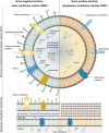
Figure 1
Figure 1
Structure and composition of bEVs secreted by Gram-negative and Gram-positive bacteria. Reproduced with permission from (28). Copyright 2022, Elsevier.
Figure 2
Figure 2
Immunoregulatory functions of extracellular vesicles. Reproduced with permission from (11). Copyright 2022, Springer Nature.
Figure 3
Figure 3
Modification strategies on OMVs during bEV vaccine production. Reproduced with permission (139). Copyright 2020, Frontiers.
Figure 4
Figure 4
Schematic preparation of CMCNP. Several methods are employed to isolate cell membrane vesicles, including repeated freeze-thaw cycles, hypotonic lysis, ultrasonic disruption, and gradient centrifugation. For optimal stability, the core of cell membrane vesicles is enveloped with specially designed antibiotic-loaded NPs or intrinsic antimicrobial NPs. To successfully prepare CMCNP, a range of techniques are utilized to ensure the thorough encapsulation of cell membrane vesicles around the NP. These techniques include membrane extrusion, ultrasonic fusion, microfluidic electroporation, and co-incubation. Reproduced with permission (183). Copyright 2022, Wiley.
Figure 5
Figure 5
Utilization of PNPs to actively target and eradicate bacteria in the treatment of sepsis. (A) Illustration depicting the structure, composition, and properties of PNPs. Quantitative analysis on selected PL membrane proteins using Western blotting (B). Flow cytometric analysis of phagocytized nanoparticles by human THP-1 cells (C). Scanning tunnel microscopy images of MRSA252 bacteria incubated with PBS (top left), uncoated nanoparticles (top right), RBCNPs (bottom left), and PNPs (bottom right) (D). The in vitro antimicrobial efficacy of Van in various forms (E). An in vivo assessment of the antimicrobial efficacy of Van measured by quantifying bacterial counts in different organs (blood, heart, lung, liver, spleen, and kidney) of mice that were infected with MRSA252. The mice were subjected to various forms and doses of Van for a period of 3 days (F). *P ≤ 0.05, **P ≤ 0.01, ***P ≤ 0.001, ****P ≤ 0.0001. Reproduced with permission (185). Copyright 2015, Springer Nature.
Figure 6
Figure 6
(A) This figure illustrates the processes of neutralizing endotoxin and inhibiting proinflammatory cytokines, which are two crucial mechanisms employed by MΦ-NPs in the treatment of sepsis. To measure the neutralization of LPS by MΦ-NPs in vitro, a fixed quantity of MΦ-NPs (0.4 mg) was introduced alongside different quantities of LPS (B). The therapeutic effects of MΦ-NPs were evaluated by administering lethal doses of E. coli-induced bacteremia (C–F). The survival curves of mice with bacteremia after treatment with MΦ-NPs (n = 10) (C). Bacterial counts in the blood, spleen, kidney, and liver within 4 hours after intraperitoneal injection of MΦ-NPs (D). Levels of pro-inflammatory cytokines, including IL-6, TNF-α, and IFN-γ, in blood and spleen samples (E, F). ns, not significant; *P < 0.05, **P < 0.01. Reproduced with permission from (189). Copyright 2017, National Academy of Sciences.

Similar articles

Cited by

References

    1. Zhang Y, Han J. Rethinking sepsis after a two-year battle with COVID-19. Cell Mol Immunol (2022) 19(11):1317–8. doi: 10.1038/s41423-022-00909-7 - DOI - PMC - PubMed
    1. Liu H, Hou S, Tian X. Risk factors of sepsis associated acute kidney injury in patients with sepsis: A meta-analysis. Intensive Care Res (2023) 3(2):163–70. doi: 10.1007/s44231-023-00034-7 - DOI
    1. Chen L, Huang Q, Zhao T, Sui L, Wang S, Xiao Z, et al. . Nanotherapies for sepsis by regulating inflammatory signals and reactive oxygen and nitrogen species: New insight for treating COVID-19. Redox Biol (2021) 45:102046. doi: 10.1016/j.redox.2021.102046 - DOI - PMC - PubMed
    1. Sun T, Wang L, Zhang H. Intestinal microbiota in sepsis. Intensive Care Res (2022) 2(1):1–7. doi: 10.1007/s44231-022-00001-8 - DOI
    1. Mithal LB, Arshad M, Swigart LR, Khanolkar A, Ahmed A, Coates BM. Mechanisms and modulation of sepsis-induced immune dysfunction in children. Pediatr Res (2022) 91(2):447–53. doi: 10.1038/s41390-021-01879-8 - DOI - PMC - PubMed

Publication types

Substances