

Depth-selective method for time-domain diffuse reflectance measurements: validation study of the dual subtraction technique
- PMID: 38420319
- PMCID: PMC10898577
- DOI: 10.1364/BOE.497671
Depth-selective method for time-domain diffuse reflectance measurements: validation study of the dual subtraction technique
Abstract
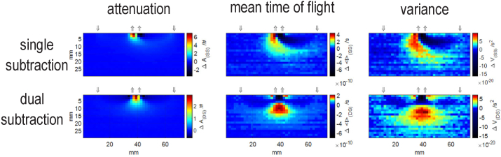
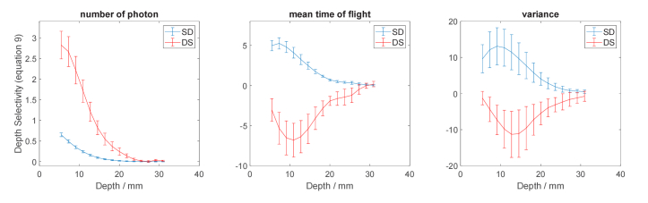
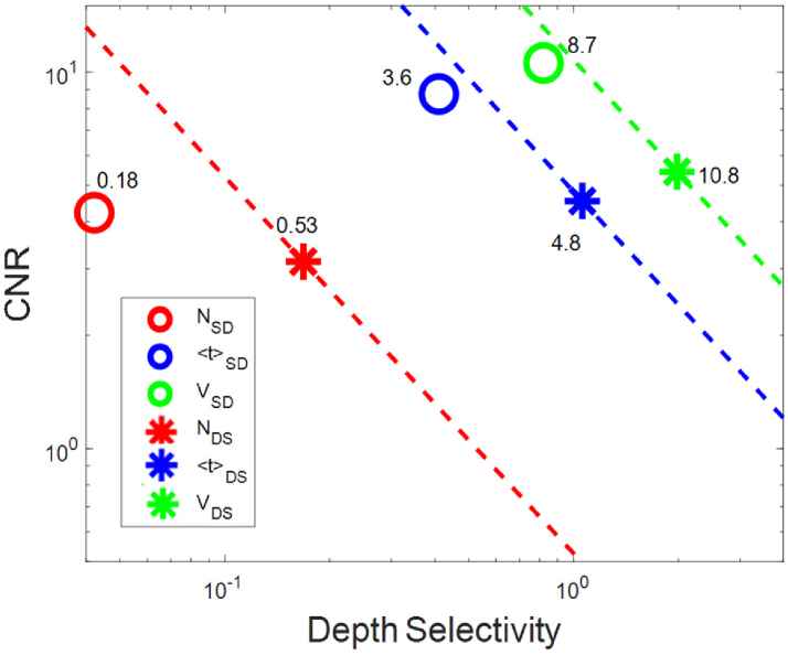
Research on the spatial distribution of sensitivity of time-domain near infrared diffuse reflectance measurement is reported in this paper. The main objective of the investigation is to validate theoretically calculated sensitivity profiles for a measurement geometry with two detectors and two sources in which sensitivity profiles of statistical moments of distributions of time of flight of photons (DTOFs) are spatially restricted to a region underneath the detectors. For this dual subtraction method, smaller sensitivities to changes appearing in the superficial layer of the medium were observed compared to the single distance and single subtraction methods. Experimental validation of this approach is based on evaluation of changes in the statistical moments of DTOFs measured on a liquid phantom with local absorption perturbations. The spatial distributions of sensitivities, depth-related sensitivity and depth selectivities were obtained from the dual subtraction method and compared with those from single distance and single subtraction approaches. Also, the contrast to noise ratio (CNR) was calculated for the dual subtraction technique and combined with depth selectivity in order to assess the overall performance (product of CNR and depth selectivity) of the method. Spatial sensitivity profiles from phantom experiments are in a good agreement with the results of theoretical studies and feature more locally restricted sensitivity volume with the point of maximal sensitivity located deeper. The highest value of overall performance was obtained experimentally for the second statistical moment in the dual subtraction method (∼10.8) surpassing that of the single distance method (∼8.7). This confirms the advantage of dual subtraction measurement geometries in the suppression of optical signals originated in the superficial layer of the medium.
© 2023 The Author(s).
Conflict of interest statement
The authors declare no conflicts of interest.
Figures


Similar articles
-
Method to improve the depth sensitivity of diffuse reflectance measurements to absorption changes in optically turbid medium.Biomed Opt Express. 2019 Sep 11;10(10):5031-5041. doi: 10.1364/BOE.10.005031. eCollection 2019 Oct 1. Biomed Opt Express. 2019. PMID: 31646028 Free PMC article.
-
Transformational change in the field of diffuse optics: From going bananas to going nuts.J Innov Opt Health Sci. 2020 Jan;13(1):1930013. doi: 10.1142/s1793545819300131. J Innov Opt Health Sci. 2020. PMID: 36340430 Free PMC article.
-
Phantom study to evaluate contrast-medium-enhanced digital subtraction mammography with a full-field indirect-detection system.Med Phys. 2010 Feb;37(2):577-89. doi: 10.1118/1.3276733. Med Phys. 2010. PMID: 20229866
-
Performance of measurands in time-domain optical brain imaging: depth selectivity versus contrast-to-noise ratio.Biomed Opt Express. 2020 Jul 16;11(8):4348-4365. doi: 10.1364/BOE.397483. eCollection 2020 Aug 1. Biomed Opt Express. 2020. PMID: 32923048 Free PMC article.
-
Two-layered blood-lipid phantom and method to determine absorption and oxygenation employing changes in moments of DTOFs.Biomed Opt Express. 2023 Jun 21;14(7):3506-3531. doi: 10.1364/BOE.492168. eCollection 2023 Jul 1. Biomed Opt Express. 2023. PMID: 37497481 Free PMC article.
Cited by
-
Spatial Sensitivity to Absorption Changes for Various Near-Infrared Spectroscopy Methods: A Compendium Review.J Innov Opt Health Sci. 2024 Jul;17(4):2430001. doi: 10.1142/s1793545824300015. Epub 2024 Feb 24. J Innov Opt Health Sci. 2024. PMID: 39267952 Free PMC article.
-
Relationship Between Signals from Cerebral near Infrared Spectroscopy Sensor Technology and Objectively Measured Cerebral Blood Volume: A Systematic Scoping Review.Sensors (Basel). 2025 Feb 3;25(3):908. doi: 10.3390/s25030908. Sensors (Basel). 2025. PMID: 39943547 Free PMC article.
-
Phase-based structured interrogation frequency-domain near-infrared spectroscopy.J Opt Soc Am A Opt Image Sci Vis. 2024 Aug 1;41(8):1500-1512. doi: 10.1364/JOSAA.523194. J Opt Soc Am A Opt Image Sci Vis. 2024. PMID: 39873575 Free PMC article.
-
Optimizing spatial accuracy in electroencephalography reconstruction through diffuse optical tomography priors in the auditory cortex.Biomed Opt Express. 2024 Jul 29;15(8):4859-4876. doi: 10.1364/BOE.531576. eCollection 2024 Aug 1. Biomed Opt Express. 2024. PMID: 39347003 Free PMC article.
References
-
- Nguyen N. T., Takakura H., Nishijo H., Ueda N., Ito S., Fujisaka M., Akaogi K., Shojaku H., “Cerebral hemodynamic responses to the sensory conflict between visual and rotary vestibular stimuli: an analysis with a multichannel near-infrared spectroscopy (NIRS) system,” Front. Hum. Neurosci. 14, 125 (2020).10.3389/fnhum.2020.00125 - DOI - PMC - PubMed
-
- Lange F., Tachtsidis I., “Clinical brain monitoring with time domain NIRS: a review and future perspectives,” Appl. Sci. 9(8), 1612 (2019).10.3390/app9081612 - DOI
-
- Chen W.-L., Wagner J., Heugel N., Sugar J., Lee Y.-W., Conant L., Malloy M., Heffernan J., Quirk B., Zinos A., “Functional near-infrared spectroscopy and its clinical application in the field of neuroscience: advances and future directions,” Front. Neurosci. 14, 724 (2020).10.3389/fnins.2020.00724 - DOI - PMC - PubMed
-
- Samaei S., Mogharari N., Borycki D., Liebert A., Kacprzak M., “New hybrid time-domain device for diffuse correlation spectroscopy and near-infrared spectroscopy for brain hemodynamic assessment,” in European Conference on Biomedical Optics (Optical Society of America, 2021), p. ES1B. 7.
LinkOut - more resources
Full Text Sources