

Dirty Mass sign of abdominal imaging: A case of silent pneumoperitoneum
- PMID: 38420345
- PMCID: PMC10899068
- DOI: 10.1016/j.radcr.2024.02.012
Dirty Mass sign of abdominal imaging: A case of silent pneumoperitoneum
Abstract
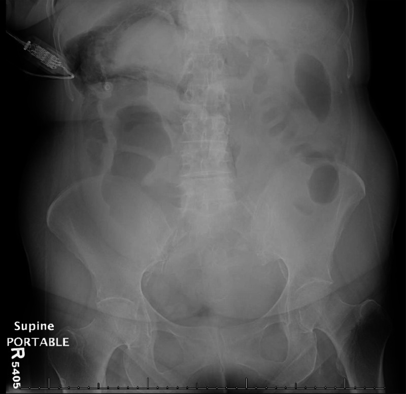
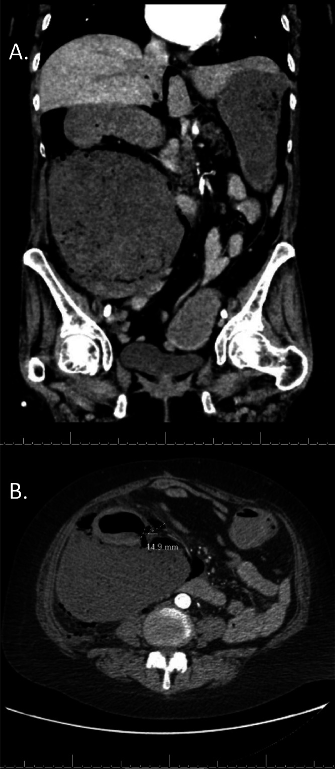
Pneumoperitoneum is a common complication after penetrating abdominal trauma, gastric ulcer, or colitis in which free air is present in the peritoneal or retroperitoneal space. Sole pneumoretroperitoneum, which refers to gas in the retroperitoneal space, is a rare entity, and when significant, results in a characteristic radiographic sign known as "Dirty Mass." Common causes include penetrating trauma or perforation of the retroperitoneal portions of the gastrointestinal tract (duodenum, ascending colon, descending colon, and rectum). Our case describes a 59-year-old female admitted for sudden onset RLQ abdominal pain with Dirty Mass sign on abdominal KUB. Early recognition of these key radiographic findings accelerates management and reduces the risk of developing complications.
Keywords: Colonic perforation; Dirty Mass; Dirty Mass sign; Pneumoretroperitoneum; Retroperitoneum.
© 2024 The Authors.
Figures
Similar articles
-
Clinical signs of retroperitoneal abscess from colonic perforation: Two case reports and literature review.Medicine (Baltimore). 2018 Nov;97(45):e13176. doi: 10.1097/MD.0000000000013176. Medicine (Baltimore). 2018. PMID: 30407351 Free PMC article. Review.
-
[Sonographical diagnosis of pneumoretroperitoneum as a result of retroperitoneal perforation].Ultraschall Med. 2007 Dec;28(6):612-21. doi: 10.1055/s-2007-963216. Epub 2007 Aug 16. Ultraschall Med. 2007. PMID: 17703376 German.
-
[New computerized tomography sign of intestinal infarction: isolated pneumoretroperitoneum or associated with pneumoperitoneum or late findings of intestinal infarction].Radiol Med. 1998 May;95(5):474-80. Radiol Med. 1998. PMID: 9687924 Italian.
-
Reappraisal of radiographic signs of pneumoperitoneum at emergency department.Am J Emerg Med. 2009 Mar;27(3):320-7. doi: 10.1016/j.ajem.2008.03.004. Am J Emerg Med. 2009. PMID: 19328377
-
Recognition of Extraperitoneal Colonic Perforation following Colonoscopy: A Review of the Literature.Case Rep Gastroenterol. 2017 May 2;11(1):256-264. doi: 10.1159/000475750. eCollection 2017 Jan-Apr. Case Rep Gastroenterol. 2017. PMID: 28559786 Free PMC article. Review.
References
-
- Saeki M., Hoshikawa Y., Miyazaki O., Nakayama F., Okamoto E., Ishikawa T. Computed tomographic analysis of colonic perforation: “Dirty mass,” a new computed tomographic finding. Emerg Radiol. 1998;5:140–145.
Publication types
LinkOut - more resources
Full Text Sources
