. 2024 Feb 10;27(3):109187.
doi: 10.1016/j.isci.2024.109187. eCollection 2024 Mar 15.

Downregulation of IRF8 in alveolar macrophages by G-CSF promotes metastatic tumor progression

Affiliations

Downregulation of IRF8 in alveolar macrophages by G-CSF promotes metastatic tumor progression

Stephanie L Tzetzo et al. iScience. .

Abstract

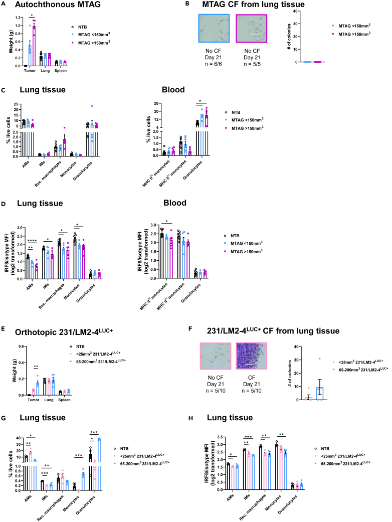
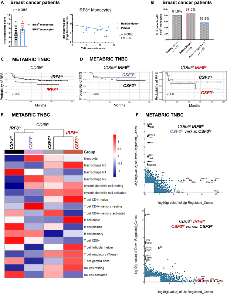
Tissue-resident macrophages (TRMs) are abundant immune cells within pre-metastatic sites, yet their functional contributions to metastasis remain incompletely understood. Here, we show that alveolar macrophages (AMs), the main TRMs of the lung, are susceptible to downregulation of the immune stimulatory transcription factor IRF8, impairing anti-metastatic activity in models of metastatic breast cancer. G-CSF is a key tumor-associated factor (TAF) that acts upon AMs to reduce IRF8 levels and facilitate metastasis. Translational relevance of IRF8 downregulation was observed among macrophage precursors in breast cancer and a CD68hiIRF8loG-CSFhi gene signature suggests poorer prognosis in triple-negative breast cancer (TNBC), a G-CSF-expressing subtype. Our data highlight the underappreciated, pro-metastatic roles of AMs in response to G-CSF and identify the contribution of IRF8-deficient AMs to metastatic burden. AMs are an attractive target of local neoadjuvant G-CSF blockade to recover anti-metastatic activity.

Keywords: Cancer; Immunology; Microenvironment.


Conflict of interest statement

M.O. has received research support from Alphageneron, AIM Therapeutics, Eli Lilly, and Pfizer. The other authors declare no competing interests.

Figures

None
Graphical abstract
Figure 1
Figure 1
IRF8 expression declines among alveolar macrophages (AMs), recruited macrophages and monocytes prior to 4T1 micro-metastasis in lung tissue (A) Mammary tumor, lung and spleen weights of BALB/c orthotopic 4T1-bearing WT mice compared to non-tumor-bearing (NTB) control tissue. (B) 4T1 micro-metastasis detection in lung tissue by staining of colony formation (CF). Scale bar: 370 μm. (C) Myeloid cell percentages within lung tissue or blood during 4T1 growth. (D) Myeloid intracellular IRF8 levels within lung tissue of NTB or 4T1-bearing mice. (E) Flow-sorted AMs have reduced Irf8 and target gene expression during 4T1 growth. (F) IRF8 levels as in (D) within blood. Data are represented as mean ± SEM. Significance was determined by Wilcoxon rank-sum tests with Holm-Bonferroni correction for pre-planned comparisons (A and B), Dunnett’s test for correction of comparisons to NTB control (C; D and F, left), Spearman correlation (D and F, right; line indicates simple linear regression), or Mann-Whitney (E; n ≥ 5 mice/group). ∗p < 0.05; ∗∗p < 0.01; ∗∗∗p < 0.001; ∗∗∗∗p < 0.0001. See also Figures S1–S3.
Figure 2
Figure 2
Macrophage IRF8 downregulation also occurs in the autochthonous MTAG model and within a mouse-human xenograft model (A) Multifocal mouse mammary tumor virus-polyomavirus middle T antigen (MTAG) tumors, lung and spleen weights compared to NTB tissue of C57BL/6 WT mice. (B) Lung tissue of MTAG mice lack micro-metastasis. Scale bar: 370 μm. (C and D) Myeloid cell percentages (C) and IRF8 levels (D) within lung tissue or blood during MTAG growth. (E) Mammary tumor, lung and spleen weights of BALB/c orthotopic 231/LM2-4LUC+-bearing SCID mice compared to NTB tissue. (F) Detection of 231/LM2-4LUC+ micro-metastasis in lung tissue. Scale bar: 370 μm. (G and H) Myeloid cell percentages (G) and IRF8 levels (H) within lung tissue during 231/LM2-4LUC+ tumor growth. Data are represented as mean ± SEM. Significance was determined by Wilcoxon rank-sum tests with Holm-Bonferroni correction for pre-planned comparisons (A and E), Mann-Whitney (B and F) or Dunnett’s test for correction of comparisons to NTB control (C, D, G, and H; n ≥ 3 mice/group). ∗p < 0.05; ∗∗p < 0.01; ∗∗∗p < 0.001; ∗∗∗∗p < 0.0001.
Figure 3
Figure 3
IRF8 expression in AMs promotes anti-metastatic activity (A) Greater spontaneous 4T1 metastatic tumor CF occurs in BALB/c IRF8 cKO (Lyz2CreIRF8fl/fl) lung tissue than IRF8fl/fl lung tissue. (B) IRF8 cKO hosts have greater 4T1 experimental lung metastasis and larger nodule sizes than IRF8fl/fl controls. (C) Schematic of control or clodronate-encapsulated liposome treatment before and during 4T1 experimental metastasis. (D and E) AM depletion via intranasal (i.n.) clodronate treatment reduces 4T1 experimental metastatic burden in IRF8 cKO hosts (D). Depletion of systemic macrophages, but not AMs, by intraperitoneal (i.p.) clodronate treatment increases 4T1 experimental metastasis in IRF8 cKO hosts (E). Representative H&E staining and images of lung metastasis displayed on the left. Scale bar: 370 μm. All data are represented as mean ± SEM. Significance was determined by Mann-Whitney (A and B) or Wilcoxon rank-sum tests with Holm-Bonferroni correction for pre-planned comparisons (D and E; n ≥ 5 mice/group). ∗p < 0.05; ∗∗p < 0.01; ∗∗∗p < 0.001. See also Figures S1–S6.
Figure 4
Figure 4
IRF8-deficient neonate liver-derived alveolar macrophages (NLDAMs) directly facilitate tumor growth (A) IRF8 cKO NLDAMs have reduced Irf8 and target gene expression (top) while increased inducible pro-tumor factor gene expression (bottom) compared to IRF8fl/fl NLDAMs. (B) Schematic of 4T1 indirect co-culture with NLDAMs or BMDMs. (C and D) IRF8 cKO NLDAMs stimulate the greatest CF compared to IRF8fl/fl controls or BMDMs (C). IRF8-transfected WT NLDAMs stimulate lower CF than IRF8-transfected IRF8 cKO NLDAMs (D). Scale bar: 370 μm. (E) Schematic of i.n. adoptive cell transfers of NLDAMs during 4T1 experimental metastasis. (F and G) IRF8 cKO NLDAM transfers augment 4T1 experimental metastasis in IRF8 cKO hosts (F). Reduced 4T1 experimental metastatic burden in IRF8 cKO hosts following transfers of IRF8-transfected WT NLDAMs than IRF8-transfected IRF8 cKO NLDAMs (G). Representative images are displayed. All data are represented as mean ± SEM. Significance was determined by Wilcoxon rank-sum tests with Holm-Bonferroni correction for pre-planned comparisons ($ indicates significantly different from all groups; n ≥ 6 mice/group). ∗p < 0.05; ∗∗p < 0.01; ∗∗∗p < 0.001; ∗∗∗∗p < 0.0001. See also Figures S7 and S8.
Figure 5
Figure 5
Mammary tumor expression of G-CSF reduces IRF8 expression in AMs and promotes lung metastasis (A) G-CSF levels within in vitro tumor-conditioned media (left) or in vivo orthotopic mammary tumors (right). (B) Triple-negative breast cancer (TNBC) cells express more G-CSF than non-TNBC cells. (C) Lung and spleen weights of NTB WT mice following local recombinant G-CSF (rG-CSF) exposure. (D and E) Myeloid cell percentages (D) and IRF8 levels (E) within lung tissue of control or rG-CSF-treated mice. (F) Schematic of local G-CSF blockade during orthotopic 4T1 growth. Tumor, lung and spleen weights from treated 4T1-bearing WT mice compared to isotype controls and baseline NTB tissue. (G) I.n. anti-G-CSF reduces lung micro-metastasis compared to isotype control treatment of 4T1-bearing mice. (H) Myeloid cell percentages within lung tissue following anti-G-CSF treatment. (I) Schematic of local G-CSF blockade during orthotopic 231/LM2-4LUC+ growth and weights as in (F). (J) I.n. blockade of human or murine G-CSF reduces lung micro-metastasis of 231/LM2-4LUC+-bearing SCID mice compared to isotype controls. (K) Myeloid cell percentages within lung tissue following human or murine G-CSF blockade. All data are represented as mean ± SEM. Significance was determined by Wilcoxon rank-sum tests with Holm-Bonferroni correction for pre-planned comparisons (A, F, I, and J), unpaired t test with Welch’s correction (B and E), Mann-Whitney (C, D and G), or Dunnett’s test for correction of comparisons to NTB control (H and K; n ≥ 5 mice/group). ∗p < 0.05; ∗∗p < 0.01; ∗∗∗p < 0.001; ∗∗∗∗p < 0.0001. See also Figure S9 and Table S1.
Figure 6
Figure 6
Local G-CSF targeting recovers anti-metastatic activity via AM- and IRF8-dependent mechanisms (A) Schematic of local neoadjuvant G-CSF blockade during orthotopic 4T1 growth compared to neoadjuvant isotype treatment. Tumor weights measured upon surgical removal. Post-surgery endpoint weights of lung or spleen. (B and C) Local neoadjuvant G-CSF blockade extends survival (B) and reduces overall metastasis (C) of WT but not IRF8 cKO mice. (D) Schematic of local neoadjuvant G-CSF blockade followed by single i.n. control or clodronate treatment during orthotopic 4T1 growth. Weights as in (A). (E and F) AM depletion via i.n. clodronate treatment does not alter post-surgery survival (E) of WT mice but negates anti-metastatic effects of local neoadjuvant G-CSF blockade (F). Representative images at post-surgery endpoint. All data are represented as mean ± SEM. Significance was determined by Wilcoxon rank-sum tests with Holm-Bonferroni correction for pre-planned comparisons (A and C), Log rank (B and E) or Mann-Whitney (D and F; n ≥ 8 mice/group). ∗p < 0.05; ∗∗p < 0.01; ∗∗∗p < 0.001; ∗∗∗∗p < 0.0001.
Figure 7
Figure 7
CD68hiIRF8hiG-CSFlo signatures within TNBC predict immune effector gene enrichment (A and B) Monocyte IRF8 levels of healthy donor or breast cancer patients were quantified by flow cytometry. Primary tumor, regional lymph node and metastasis (TNM) composite scores trend higher in patients with IRF8lo monocytes (A, left) and inversely correlate with IRF8 levels (A, right). Downward trend in the percentage of patients with IRF8hi monocytes as breast cancer stage advances (B). (C) Relapse-free survival (RFS) of METABRIC TNBC patients (n = 223) with a CD68hiIRF8hi signature trends toward more favorable outcome than CD68hiIRF8lo. (D) High G-CSF (encoded by CSF3) predicts a trend in poorer outcome of the TNBC CD68hiIRF8lo signature (right), but not in the CD68hiIRF8hi signature (left). (E and F) Immune enrichment (E) and MR analyses (F) based on differential IRF8 and G-CSF expression in TNBC CD68hi signatures. Data are represented as mean ± SEM. Significance was determined by unpaired t test with Welch’s correction (A, left), Spearman correlation (A, right; line indicates simple linear regression) or Log rank test (C and D). ∗∗p < 0.01. Trend analyses were determined by one-sided Cochrane-Armitage test (B; p = 0.03402; n ≥ 8 patients/group). See also Figures S9, S10 and Table S2.

References

    1. Anderson R.L., Balasas T., Callaghan J., Coombes R.C., Evans J., Hall J.A., Kinrade S., Jones D., Jones P.S., Jones R., et al. A framework for the development of effective anti-metastatic agents. Nat. Rev. Clin. Oncol. 2019;16:185–204. doi: 10.1038/s41571-018-0134-8. - DOI - PMC - PubMed
    1. Ganesh K., Massagué J. Targeting metastatic cancer. Nat. Med. 2021;27:34–44. doi: 10.1038/s41591-020-01195-4. - DOI - PMC - PubMed
    1. Liu Y., Cao X. Characteristics and Significance of the Pre-metastatic Niche. Cancer Cell. 2016;30:668–681. doi: 10.1016/j.ccell.2016.09.011. - DOI - PubMed
    1. Correia A.L. Locally sourced: site-specific immune barriers to metastasis. Nat. Rev. Immunol. 2023;23:522–538. doi: 10.1038/s41577-023-00836-2. - DOI - PMC - PubMed
    1. Patras L., Shaashua L., Matei I., Lyden D. Immune determinants of the pre-metastatic niche. Cancer Cell. 2023;41:546–572. doi: 10.1016/j.ccell.2023.02.018. - DOI - PMC - PubMed

LinkOut - more resources