. 2024 Feb 5;27(3):109125.
doi: 10.1016/j.isci.2024.109125. eCollection 2024 Mar 15.

YAP-mediated GPER signaling impedes proliferation and survival of prostate epithelium in benign prostatic hyperplasia

Affiliations

YAP-mediated GPER signaling impedes proliferation and survival of prostate epithelium in benign prostatic hyperplasia

Zhifu Liu et al. iScience. .

Abstract

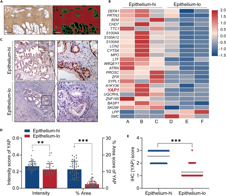
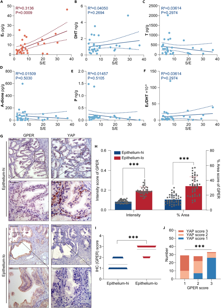
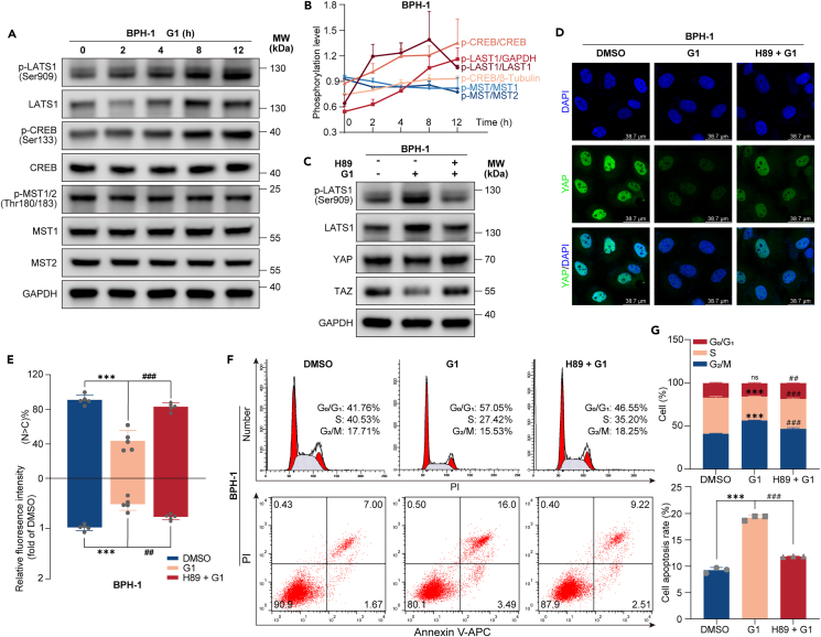
Benign prostatic hyperplasia (BPH) occurs when there is an imbalance between the proliferation and death of prostate cells, which is regulated tightly by estrogen signaling. However, the role of G protein-coupled estrogen receptor (GPER) in prostate cell survival remains ambiguous. In this study, we observed that prostates with epithelial hyperplasia showed increased yes-associated protein 1 (YAP) expression and decreased levels of estrogen and GPER. Blocking YAP through genetic or drug interventions led to reduced proliferation and increased apoptosis in the prostate epithelial cells. Interestingly, GPER agonists produced similar effects. GPER activation enhanced the phosphorylation and degradation of YAP, which was crucial for suppressing cell proliferation and survival. The Gαs/cAMP/PKA/LATS pathway, downstream of GPER, transmitted signals that facilitated YAP inhibition. This study investigated the interaction between GPER and YAP in the prostate epithelial cells and its contribution to BPH development. It lays the groundwork for future research on developing BPH treatments.

Keywords: Biochemistry; Cell biology; Molecular biology; Prostate disease.


Conflict of interest statement

The authors declare no competing interests.

Figures

None
Graphical abstract
Figure 1
Figure 1
YAP expression is elevated in epithelium-rich BPH group (A) Left: Representative staining of α-SMA in BPH tissue. Right: Analysis of the same tissue specimen showing epithelial and stromal components using the Strata Quest Histo system (stroma in red, epithelia in green; Scale bars: 100 μm). (B) Heatmap illustrating the differentially expressed proteins in BPH tissues from Epithelium-hi compared to Epithelium-lo, including YAP. (C) Representative immunohistochemical staining for YAP in Epithelium-hi and Epithelium-lo BPH tissues, demonstrating distinct expression patterns (Scale bars: 100 μm [left]; 25 μm [right]). (D) Scatterplot depicting the variation in YAP staining intensity and area within epithelial cells between the Epithelium-hi and Epithelium-lo groups. (E) Comparative scores of epithelial YAP expression in the two groups (0 = lowest, 3 = highest), showing significantly higher YAP expression in the epithelial cells of the Epithelium-hi group. Data were presented as mean ± SD (n = 45). Significant difference was determined by two-tailed unpaired t test (D) or Mann-Whitney test (E) (∗∗p < 0.01, ∗∗∗p < 0.001).
Figure 2
Figure 2
Restraint of YAP inhibits cell proliferation and induces cell apoptosis (A‒F) BPH-1 and RWPE-1 cell lines were subjected to either DMSO treatment or indicated concentrations of verteporfin. (A and B): Cell growth dynamics of both cell lines were monitored through CCK-8 assays conducted at 24-h intervals. (C and D): After 48 h, FCM analysis determined the distribution of cells across different cell cycle phases, with the histograms representing the proportion of cells in each phase. (E and F): Cell apoptosis was assessed after 48 h using Annexin V-FITC/PI staining and FACS, with the histograms illustrating the apoptosis rates. (G) The efficacy of siRNAs targeting YAP was evaluated using RT-qPCR, with GAPDH serving as the endogenous control. (H and I) Post-RNAi knockdown of YAP in both cell lines, cell growth curves were charted using CCK-8 assays at 24-h intervals. (J and K) Western blot analysis was conducted to measure the relative expression levels of YAP, CDK4/6, cyclin D1, Bax, and Bcl-2 following either verteporfin treatment or RNAi intervention. β-Tubulin was used as a loading control. All blots shown are representative of three experimental replicates. Data are expressed as mean ± SD from a minimum of three independent experiments. Significant difference was determined by one way ANOVA followed by a Tukey’s multiple comparison tests (∗p < 0.05, ∗∗p < 0.01, ∗∗∗p < 0.001).
Figure 3
Figure 3
YAP expression is negatively related to tissue estrogen and GPER level (A‒F) These scatterplots display the levels of various hormones—17β-estradiol (E2), dihydrotestosterone (DHT), testosterone (T), androstenedione (A-dione), and progesterone (P) —in BPH specimens characterized by S/E ratios. Notably, a higher S/E ratio was positively correlated with increased levels of E2 in the tissues (R = 0.3136, p < 0.001, n = 32). However, no significant correlation was observed between the S/E ratio and the concentrations of other hormones studied. (G) This panel shows consecutive histological sections of epithelial cells from the Epithelium-hi and Epithelium-lo groups, stained to reveal the expression of GPER and YAP, respectively (Scale bars: 100 μm [up]; 25 μm [down]). (H) A scatterplot illustrating the scores for epithelial GPER staining intensity and area in the Epithelium-hi and Epithelium-lo groups (n = 45). (I) Comparative scores of epithelial GPER expression in the two groups (0 = lowest, 3 = highest) (n = 45). (J) A negative correlation was observed between the expression of YAP and GPER in the epithelial cells. Data are expressed as mean ± SD. Significant difference was determined by Pearson’s correlation analysis (A–F), two-tailed unpaired t test (H), Mann-Whitney test (E), or Chi-square test (J) (∗∗∗p < 0.001).
Figure 4
Figure 4
Activation of GPER impedes the proliferation and survival of prostate epithelial cells (A and B) Growth curves for BPH-1 and RWPE-1 cell lines were established following treatment with G1, E2, or OHT at specified concentrations, using CCK-8 assays conducted every 24 h. (C and D) Histograms illustrating cell viability on a daily basis with representative data. (E and F) FCM assessed the distribution of cells across various cell cycle phases after 48 h of treatment with G1 (1 μM) and E2 (10 nM). The histograms depict the proportion of cells in each specific phase. (G and H) Cell apoptosis was evaluated using Annexin V-FITC/PI staining FACS following GPER activation by G1 and E2 treatments, with histograms illustrating the apoptosis rates. (I) Pre-treatment with GPER blocker G15 (5 μM) for 1 h mitigated the G1 and E2-induced inhibition of cell proliferation and survival. Cell survival rates were quantified using CCK-8 assays. (J) GPER blockade via siRNA reduced the suppression of cell survival caused by G1, determined using CCK-8 assays. (K) Western blot analysis was performed to determine the relative expression levels of CDK4/6, cyclin D1, Bax, and Bcl-2 post-treatment with G1 and E2. β-Tubulin was used as the loading control. (L) The relative expression of aforementioned proteins was analyzed by western blot after pre-treatment with G15. β-Tubulin served as the loading control. All blots shown are representative of three experimental replicates. Data are expressed as mean ± SD from a minimum of three independent experiments. Significant difference was determined by one way ANOVA followed by a Tukey’s multiple comparison tests (∗p < 0.05, ∗∗p < 0.01, ∗∗∗p < 0.001).
Figure 5
Figure 5
GPER acts via inhibition of YAP (A) GPER activation led to the phosphorylation of YAP in BPH-1 cells treated with 1 μM G1, 10 nM E2, or 5 μM OHT. Immunoblotting was utilized to assess the relative expression levels of p-YAP/YAP, p-TAZ/TAZ, with GAPDH serving as the loading control. (B and C) GPER mediated YAP phosphorylation. GPER was inhibited by 5 μM G15 pre-treatment (B) or siRNAs (C). Following this, BPH-1 cells were treated with G1, and immunoblotting was conducted to analyze the phosphorylation levels. (D) Activation of GPER suppressed the expression of YAP-induced target genes. BPH-1 cells treated with G1 or E2 for 24 h were subjected to quantitative PCR to measure mRNA levels of specific target genes. (E and F) G1 suppressed YAP’s expression and nuclear localization. BPH-1 cells were treated with G1, with or without G15 pre-treatment. Immunofluorescence staining for YAP was performed, and quantifications of YAP’s subcellular localization and staining intensity were done based on 5 randomly selected fields. N indicates nuclear localization; C indicates cytoplasmic. (G) GPER activation hindered the nuclear translocation of YAP and its interaction with TEAD1 in BPH-1 cells. After 24 h of G1 treatment, cells were subjected to immunoprecipitation using an anti-YAP antibody, followed by detection of coimmunoprecipitated TEAD1. (H‒M) YAP was required for GPER to impede the proliferation and survival of prostate epithelial cells. (H): Overexpression of YAP significantly diminished the impact of G1 and E2 on cell proliferation, as determined by the CCK-8 assay. (I and J): FCM was used to evaluate the distribution of cells across different cell cycle phases, with histograms indicating the proportion of cells in each phase. (K and L): Cell apoptosis was assessed using Annexin V-FITC/PI staining FACS, with histograms showing apoptosis rates. (M): Immunoblotting was performed to analyze the relative expression of YAP, CDK4/6, cyclin D1, Bax, and Bcl-2 following treatment with G1 and E2, both with and without YAP overexpression. β-Tubulin was used as the loading control. All blots shown are representative of three experimental replicates. Data are expressed as mean ± SD from a minimum of three independent experiments. Significant difference was determined by one way ANOVA followed by a Tukey’s multiple comparison tests (D and F) and two-tailed unpaired t test (H, J, and L) (∗p < 0.05, ∗∗p < 0.01, ∗∗∗p < 0.001, ns for non-significant).
Figure 6
Figure 6
GPER inhibits YAP through the Gαs/cAMP/PKA pathway and LATS1 activation (A and B) Treatment of BPH-1 cells with 1 μM G1 resulted in an elevation of phosphorylation levels for CREB and LATS1, whereas the phosphorylation levels of MST1/2 were either unchanged or exhibited a slight decrease. The cells were treated and collected at 2, 4, 8, and 12 h for immunoblotting to detect the phosphorylation levels of LATS, CREB, and MST1/2. The line chart depicts the time-dependent quantification of phosphorylation levels for CREB, LATS1, and MST1/2. (C) PKA was required for G1 to inhibit YAP expression. BPH-1 cells pre-treated with 10 μM PKA inhibitor H89 for 1 h underwent immunoblotting to assess the phosphorylation level of LATS1 and the expression levels of YAP and TAZ. (D and E) Pre-treatment with H89 mitigated the G1-induced reduction in YAP expression and its nuclear localization. BPH-1 cells were treated with G1, with or without H89 pre-treatment, followed by immunofluorescence staining for YAP. Quantifications of YAP’s subcellular localization and staining intensity were conducted from 5 randomly selected fields. N indicates nuclear localization; C indicates cytoplasmic. (F and G) Blockade of PKA activity by pre-treatment with 10 μM H89 for 1 h counteracted the G1-induced cell-cycle arrest (upper) and apoptosis (lower). Histograms display the proportion of cells in each cell cycle phase and the apoptosis rates. All blots shown are representative of three experimental replicates. Data are expressed as mean ± SD from a minimum of three independent experiments. Significant difference was determined by one way ANOVA followed by a Tukey’s multiple comparison tests (∗∗∗p < 0.001 compared with DMSO treatment; ##p < 0.01, ###p < 0.001 compared with G1 treatment, ns for non-significant).

Similar articles

Cited by

References

    1. Chughtai B., Forde J.C., Thomas D.D.M., Laor L., Hossack T., Woo H.H., Te A.E., Kaplan S.A. Benign prostatic hyperplasia. Nat. Rev. Dis. Primers. 2016;2:16031. doi: 10.1038/nrdp.2016.31. - DOI - PubMed
    1. Roehrborn C.G. Pathology of benign prostatic hyperplasia. Int. J. Impot. Res. 2008;20:S11–S18. doi: 10.1038/ijir.2008.55. - DOI - PubMed
    1. Ho C.K.M., Habib F.K. Estrogen and androgen signaling in the pathogenesis of BPH. Nat. Rev. Urol. 2011;8:29–41. doi: 10.1038/nrurol.2010.207. - DOI - PubMed
    1. Ho C.K.M., Nanda J., Chapman K.E., Habib F.K. Oestrogen and benign prostatic hyperplasia: effects on stromal cell proliferation and local formation from androgen. J. Endocrinol. 2008;197:483–491. doi: 10.1677/JOE-07-0470. - DOI - PubMed
    1. Zhang Z., Duan L., Du X., Ma H., Park I., Lee C., Zhang J., Shi J. The proliferative effect of estradiol on human prostate stromal cells is mediated through activation of ERK. Prostate. 2008;68:508–516. doi: 10.1002/pros.20722. - DOI - PubMed