

Placental Pathology Contributes to Impaired Volumetric Brain Development in Neonates With Congenital Heart Disease
- PMID: 38420785
- PMCID: PMC10944035
- DOI: 10.1161/JAHA.123.033189
Placental Pathology Contributes to Impaired Volumetric Brain Development in Neonates With Congenital Heart Disease
Abstract
Background: Neonates with congenital heart disease are at risk for impaired brain development in utero, predisposing children to postnatal brain injury and adverse long-term neurodevelopmental outcomes. Given the vital role of the placenta in fetal growth, we assessed the incidence of placental pathology in fetal congenital heart disease and explored its association with total and regional brain volumes, gyrification, and brain injury after birth.
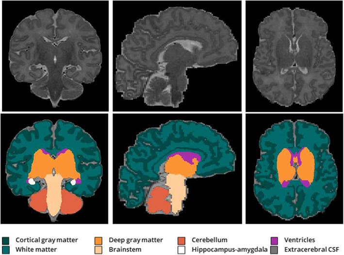
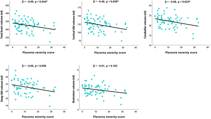
Methods and results: Placentas from 96 term singleton pregnancies with severe fetal congenital heart disease were prospectively analyzed for macroscopic and microscopic pathology. We applied a placental pathology severity score to relate placental abnormalities to neurological outcome. Postnatal, presurgical magnetic resonance imaging was used to analyze brain volumes, gyrification, and brain injuries. Placental analyses revealed the following abnormalities: maternal vascular malperfusion lesions in 46%, nucleated red blood cells in 37%, chronic inflammatory lesions in 35%, delayed maturation in 30%, and placental weight below the 10th percentile in 28%. Severity of placental pathology was negatively correlated with cortical gray matter, deep gray matter, brainstem, cerebellar, and total brain volumes (r=-0.25 to -0.31, all P<0.05). When correcting for postmenstrual age at magnetic resonance imaging in linear regression, this association remained significant for cortical gray matter, cerebellar, and total brain volume (adjusted R2=0.25-0.47, all P<0.05).
Conclusions: Placental pathology occurs frequently in neonates with severe congenital heart disease and may contribute to impaired brain development, indicated by the association between placental pathology severity and reductions in postnatal cortical, cerebellar, and total brain volumes.
Keywords: brain development; congenital heart disease; fetus; magnetic resonance imaging; neonate; neuroplacentology; placenta.
Figures
Comment in
-
Placenta-Heart-Brain Connection in Congenital Heart Disease.J Am Heart Assoc. 2024 Mar 5;13(5):e033875. doi: 10.1161/JAHA.124.033875. Epub 2024 Feb 29. J Am Heart Assoc. 2024. PMID: 38420776 Free PMC article. No abstract available.
References
-
- Bonthrone AF, Stegeman R, Feldmann M, Claessens NHP, Nijman M, Jansen NJG, Nijman J, Groenendaal F, de Vries LS, Benders MJNL, et al. Risk factors for perioperative brain lesions in infants with congenital heart disease: a European collaboration. Stroke. 2022;53:3652–3661. doi: 10.1161/STROKEAHA.122.039492 - DOI - PMC - PubMed
-
- Sun L, Macgowan CK, Sled JG, Yoo SJ, Manlhiot C, Porayette P, Grosse‐Wortmann L, Jaeggi E, McCrindle BW, Kingdom J, et al. Reduced fetal cerebral oxygen consumption is associated with smaller brain size in fetuses with congenital heart disease. Circulation. 2015;131:1313–1323. doi: 10.1161/CIRCULATIONAHA.114.013051 - DOI - PMC - PubMed
MeSH terms
LinkOut - more resources
Full Text Sources
Medical
