. 2024 Feb;18(1):69-86.
doi: 10.1080/17435390.2024.2314473. Epub 2024 Feb 29.

Potent lung tumor promotion by inhaled MWCNT

Affiliations

Potent lung tumor promotion by inhaled MWCNT

Dale W Porter et al. Nanotoxicology. 2024 Feb.

Abstract

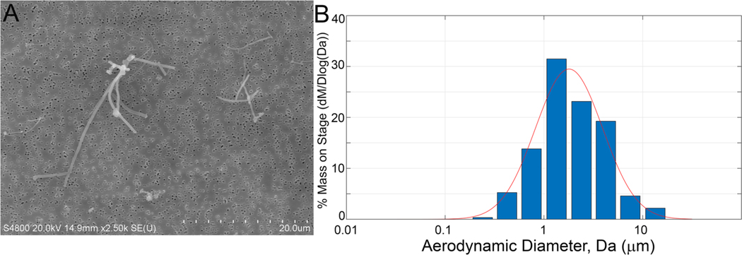
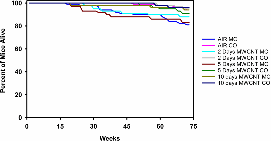
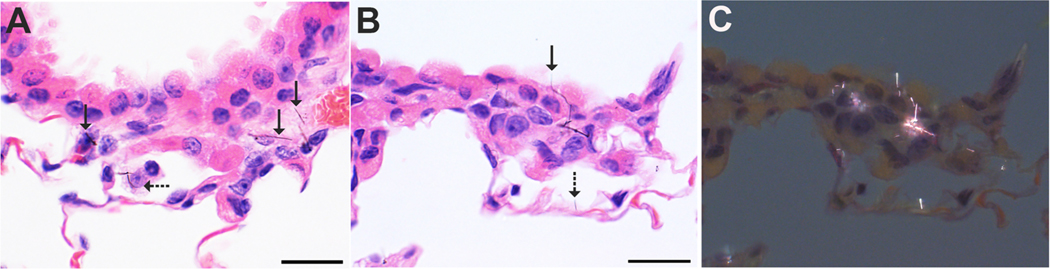
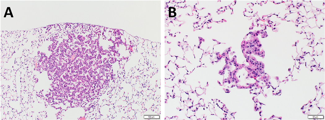
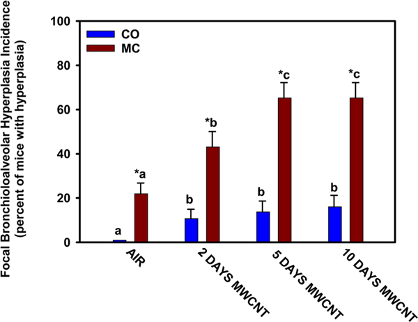
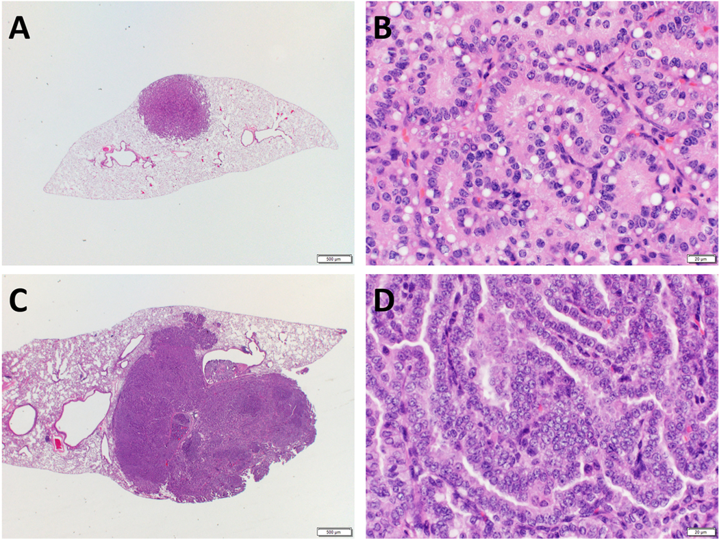
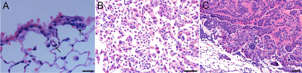
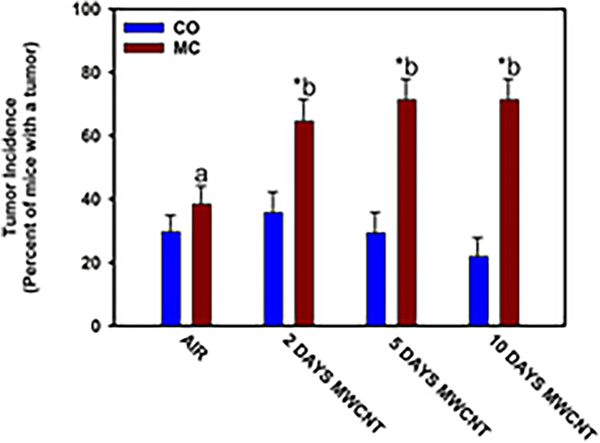
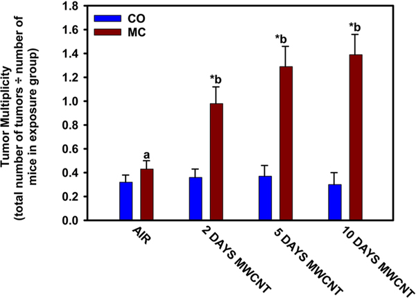
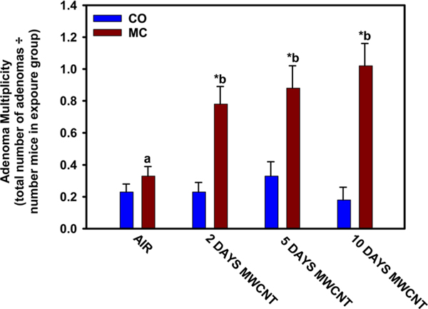
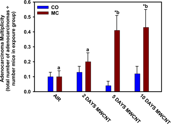
In the lung, carcinogenesis is a multi-stage process that includes initiation by a genotoxic agent, promotion that expands the population of cells with damaged DNA to form a tumor, and progression from benign to malignant neoplasms. We have previously shown that Mitsui-7, a long and rigid multi-walled carbon nanotube (MWCNT), promotes pulmonary carcinogenesis in a mouse model. To investigate the potential exposure threshold and dose-response for tumor promotion by this MWCNT, 3-methylcholanthrene (MC) initiated (10 μg/g, i.p., once) or vehicle (corn oil) treated B6C3F1 mice were exposed by inhalation to filtered air or MWCNT (5 mg/m3) for 5 h/day for 0, 2, 5, or 10 days and were followed for 17 months post-exposure for evidence of lung tumors. Pulmonary neoplasia incidence in MC-initiated mice significantly increased with each MWCNT exposure duration. Exposure to either MC or MWCNT alone did not affect pulmonary neoplasia incidence compared with vehicle controls. Lung tumor multiplicity in MC-initiated mice also significantly increased with each MWCNT exposure duration. Thus, a significantly higher lung tumor multiplicity was observed after a 10-day MWCNT exposure than following a 2-day exposure. Both bronchioloalveolar adenoma and bronchioloalveolar adenocarcinoma multiplicity in MC-initiated mice were significantly increased following 5- and 10-day MWCNT exposure, while a 2-day MWCNT exposure in MC-initiated mice significantly increased the multiplicity of adenomas but not adenocarcinomas. In this study, even the lowest MWCNT exposure promoted lung tumors in MC-initiated mice. Our findings indicate that exposure to this MWCNT strongly promotes pulmonary carcinogenesis.

Keywords: Multi-walled carbon nanotubes; inhalation exposure; lung cancer; mice; tumor promoter.


Conflict of interest statement

Disclosure statement

No potential conflict of interest was reported by the author(s).

Figures

Figure 1.
Figure 1.
MWCNT aerosol characterization. Panel A. Field emission scanning electron microscope image showing the morphology of MWCNT aerosol samples from our exposure chamber. Panel B. Mass size distribution of the MWCNT aerosol in the exposure chamber (M = mass concentration and Dae = aerodynamic diameter). The distribution has a MMAD = 1.7 μm and a GSD of 3.0.
Figure 2.
Figure 2.
Survival curve. Depicts the percent of mice alive in each treatment group throughout the duration of the study.
Figure 3.
Figure 3.
Panel A. MWCNT persisting in the bronchioloalveolar junction in an H&E section of lung from a representative mouse exposed to MWCNT for 5 days. MWCNT are located within the peribronchiolar interstitium (solid arrows) and in an alveolar macrophage (dashed arrow). Panel B. MWCNT sometimes extend beyond the cytoplasmic margins (solid arrow) as in the bronchiolar epithelium of this mouse and sometimes projected from alveolar septa (dashed arrow). Panel C. This polarized light microscopic image from the same bronchioloalveolar region shown in B demonstrates the numerous MWCNT that are not easily seen with standard light microscopy. Scale bar = 20 microns.
Figure 4.
Figure 4.
Hyperplasia. Representative photomicrographs of H&E-stained sections of lung from mice exposed to MWCNT. Focal bronchioloalveolar hyperplasia: Marked hyperplasia (Panel A) is considered a preneoplastic lesion consisting of >20 contiguous alveolar walls being affected. Minimal hyperplasia is seen in Panel B.
Figure 5.
Figure 5.
Incidence of focal bronchioloalveolar hyperplasia. All mice received a single dose of either 3-methylcholanthrene (MC: 10 μg/g bw, i.p.) or corn oil vehicle (CO). One week after receiving MC or CO, mice were exposed by whole-body inhalation to MWCNT (5 mg/m3, 5 h/day) or filtered air for 2, 5, or 10days. Mice were euthanized 17months after exposure. CO-treated mice exposed to air for 2, 5, or 10days were pooled because statistical analyses indicated no significant difference (p > 0.05) between exposure times. Similarly, the number of days of air exposure had no significant effect (p > 0.05) on MC-treated mice. Thus, MC-treated mice exposed to air for 2, 5, or 10days were also pooled. At each exposure time, an asterisk (*) indicates that the MC-treated group (MC) was significantly higher (p < 0.05) than the corresponding corn oil-treated (CO) group. For MC-exposed mice, bars with different letters are significantly different (p < 0.05).
Figure 6.
Figure 6.
Photomicrographs of bronchioloalveolar adenoma and bronchioloalveolar adenocarcinoma. Representative photomicrographs of H&E-stained sections of lung tumors from mice exposed to MWCNT. Bronchioloalveolar adenoma (Panels A and B): the adenoma is expansive and well-circumscribed, and the neoplastic cells form irregular papillary structures (Panel B). Bronchioloalveolar adenocarcinoma (Panels C and D): the adenocarcinoma is invasive and extends into the surrounding lung parenchyma. Neoplastic cells show increased cellular atypia with higher nuclear-to-cytoplasmic ratio.
Figure 7.
Figure 7.
Persistent Histiocytic Infiltration. In Panel A, a partially polarized image of histiocytic infiltration (alveolar histiocytosis) (solid arrow) and interstitial hypercellularity (dashed arrow) associated with persistent MWCNT (bar = 20 microns) are shown. Panel B is a light microscopic image of diffuse histiocytic infiltration associated with focal hyperplasia (bar = 50 microns). Panel C shows a light microscopic image of tumor-associated predominantly histiocytic infiltrates (asterisks).
Figure 8.
Figure 8.
Percent Tumor Incidence. All mice received a single dose of either 3-methylcholanthrene (MC: 10 μg/g bw, i.p.) or corn oil vehicle (CO). One week after receiving MC or CO, mice were exposed by whole-body inhalation to MWCNT (5 mg/m3, 5 h/day) or filtered air for 2, 5, or 10 days. Mice were euthanized 17 months after exposure. CO-treated mice exposed to air for 2, 5, or 10 days were pooled because statistical analyses indicated no significant difference (p > 0.05) between exposure times. Similarly, the number of days of air exposure had no significant effect (p > 0.05) on MC-treated mice. Thus, MC-treated mice exposed to air for 2, 5, or 10 days were also pooled. At each exposure time, an asterisk (*) indicates that the MC-treated group was significantly higher (p < 0.05) than the corn oil-treated group. For MC-exposed mice, bars with different letters are significantly different (p < 0.05).
Figure 9.
Figure 9.
Tumor Multiplicity. All mice received a single dose of either 3-methylcholanthrene (MC: 10 μg/g bw, i.p.) or corn oil vehicle (CO). One week after receiving MC or CO, mice were exposed by whole-body inhalation to MWCNT (5 mg/m3, 5 h/day) or filtered air for 2, 5, or 10 days. Mice were euthanized 17 months after exposure. CO-treated mice exposed to air for 2, 5, or 10 days were pooled because statistical analyses indicated no significant difference (p > 0.05) between exposure times. Similarly, the number of days of air exposure had no significant effect (p > 0.05) on MC-treated mice. Thus, MC-treated mice exposed to air for 2-, 5-, or 10-days were also pooled. At each exposure time, an asterisk (*) indicates that the MC-treated group was significantly higher (p < 0.05) than the corn oil-treated group. For MC-exposed mice, bars with different letters are significantly different (p < 0.05).
Figure 10.
Figure 10.
Adenoma multiplicity. All mice received a single dose of either 3-methylcholanthrene (MC: 10 μg/g bw, i.p.) or corn oil vehicle (CO). One week after receiving MC or CO, mice were exposed by whole-body inhalation to MWCNT (5 mg/m3, 5 h/day) or filtered air for 2, 5, or 10 days. Mice were euthanized 17 months after exposure. CO-treated mice exposed to air for 2, 5, or 10 days were pooled because statistical analyses indicated no significant difference (p > 0.05) between exposure times. Similarly, the number of days of air exposure had no significant effect (p > 0.05) on MC-treated mice. Thus, MC-treated mice exposed to air for 2, 5, or 10 days were also pooled. At each exposure time, an asterisk (*) indicates that the MC-treated group was significantly higher (p < 0.05) than the corn oil-treated group. For MC-exposed mice, bars with different letters are significantly different (p < 0.05).
Figure 11.
Figure 11.
Adenocarcinoma multiplicity. All mice received a single dose of either 3-methylcholanthrene (MC: 10 μg/g bw, i.p.) or corn oil vehicle (CO). One week after receiving MC or CO, mice were exposed by whole-body inhalation to MWCNT (5 mg/m3, 5 h/day) or filtered air for 2, 5, or 10 days. Mice were euthanized 17 months after exposure. CO-treated mice exposed to air for 2, 5, or 10 days were pooled because statistical analyses indicated no significant difference (p > 0.05) between exposure times. Similarly, the number of days of air exposure had no significant effect (p > 0.05) on MC-treated mice. Thus, MC-treated mice exposed to air for 2, 5, or 10 days were also pooled. At each exposure time, an asterisk (*) indicates that the MC-treated group was significantly higher (p < 0.05) than the corn oil-treated group. For MC-exposed mice, bars with different letters are significantly different (p < 0.05).

Similar articles

References

    1. Barbarino M, and Giordano A. 2021. “Assessment of the Carcinogenicity of Carbon Nanotubes in the Respiratory System.” Cancers 13 (6): 1318. 10.3390/cancers13061318 - DOI - PMC - PubMed
    1. Barra V, and Fachinetti D. 2018. “The Dark Side of Centromeres: Types, Causes and Consequences of Structural Abnormalities Implicating Centromeric DNA.” Nature Communications 9 (1): 4340. 10.1038/s41467-018-06545-y - DOI - PMC - PubMed
    1. Barrett JC 1993. “Mechanisms of Multistep Carcinogenesis and Carcinogen Risk Assessment.” Environmental Health Perspectives 100: 9–20. 10.1289/ehp.931009 - DOI - PMC - PubMed
    1. Barthel Hélène, Darne Christian, Gaté Laurent, Visvikis Athanase, and Seidel Carole. 2021. “Continuous Long-Term Exposure to Low Concentrations of MWCNTS Induces an Epithelial-Mesenchymal Transition in BEAS-2B Cells.” Nanomaterials (Basel, Switzerland) 11 (7): 1742. 10.3390/nano11071742 - DOI - PMC - PubMed
    1. Bauer AK, and Dwyer-Nield LD. 2021. “Two-Stage 3-Methylcholanthrene and Butylated Hydroxytoluene-Induced Lung Carcinogenesis in Mice.” Methods in Cell Biology 163: 153–173. 10.1016/bs.mcb.2020.07.003 - DOI - PubMed

Publication types

LinkOut - more resources