. 2024 Feb;30(2):e14551.
doi: 10.1111/cns.14551.

Absence in CX3CR1 receptor signaling promotes post-ischemic stroke cognitive function recovery through suppressed microglial pyroptosis in mice

Affiliations

Absence in CX3CR1 receptor signaling promotes post-ischemic stroke cognitive function recovery through suppressed microglial pyroptosis in mice

Yangyang Ge et al. CNS Neurosci Ther. 2024 Feb.

Abstract

Background: Post-stroke cognitive impairment (PSCI) is a major source of morbidity and mortality after stroke, but the pathological mechanisms remain unclear. Previous studies have demonstrated that the CX3CR1 receptor plays a crucial role in maintaining an early protective microenvironment after stroke, but whether it persistently influences cognitive dysfunction in the chronic phase requires further investigation.

Methods: Mouse was used to establish a middle cerebral artery occlusion (MCAO)/reperfusion model to study PSCI. Cognitive function was assessed by the Morris water maze (MWM) and the novel object recognition test. Neurogenesis was assessed by immunofluorescence staining with Nestin+ /Ki67+ and DCX+ /BrdU+ double-positive cells. The cerebral damage was monitored by [18 F]-DPA-714 positron emission tomography, Nissel, and TTC staining. The pyroptosis was histologically, biochemically, and electron microscopically examined.

Results: Upon MCAO, at 28 to 35 days, CX3CR1 knockout (CX3CR1-/- ) mice had better cognitive behavioral performance both in MWM and novel object recognition test than their CX3CR1+/- counterparts. Upon MCAO, at 7 days, CX3CR1-/- mice increased the numbers of Nestin+ /Ki67+ and DCX+ /BrdU+ cells, and meanwhile it decreased the protein expression of GSDMD, NLRP3 inflammasome subunit, caspase-1, mature IL-1β/IL-18, and p-P65 in the hippocampus as compared with CX3CR1+/- mice. In addition, CX3CR1-/- mice could reverse infarct volume in the hippocampus region post-stroke.

Conclusion: Our study demonstrated that CX3CR1 gene deletion was beneficial to PSCI recovery. The mechanism might lie in inhibited pyroptosis and enhanced neurogenesis. CX3CR1 receptor may serve as a therapeutic target for improving the PSCI.

Keywords: CX3C chemokine receptor 1; cognitive dysfunction; ischemic stroke; microglia.


Conflict of interest statement

The authors declare no conflicts of interest.

Figures
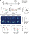
FIGURE 1
FIGURE 1
CX3CR1−/− mice enhanced the post‐ischemic stroke cognitive functional recovery. (A) Schematic showing the time of MCAO and behavior tests. CX3CR1 deficiency enhanced sensorimotor function recovery in both the mNSS test (B) and corner test (C). The novel object recognition test (D) and Morris water maze (E‐H) were employed to monitor the cognitive impairment. The behavior assessments were performed at 0, 1, 3, 5, 7, 14, 28, and 35 days after MCAO. N = 12–18. All data are shown as the mean ± SEM. *p < 0.05, **p < 0.01, ***p < 0.001 versus Sham+CX3CR1+/− group; # p < 0.05, ## p < 0.01, ### p < 0.001 versus MCAO+CX3CR1+/− group.
FIGURE 2
FIGURE 2
CX3CR1−/− mice promoted adult hippocampal neurogenesis (AHN) after ischemic stroke. (A) Representative images showing the brain sections immunostained with Nestin (green), a specific marker of NSPCs, and the endogenous marker of cell proliferation Ki67 (red) in the ipsilateral hippocampal regions at 7 days after ischemic stroke in mice. (scale bar:200 μm and 50 μm;) (B) The number of Nestin+/Ki67+ cells in CA1 and DG sub‐regions was quantified and analyzed. (C) Representative images showing the brain sections immunostained with DCX (green) and BrdU (red) in the ipsilateral hippocampal regions at 7 days after ischemic stroke in mice. (scale bar:100 μm and 20 μm) (D) The number of DCX+/Brdu+ cells in CA1 and DG were quantified and analyzed. N = 6. All the data are shown as the mean ± SEM. *p < 0.05, ***p < 0.001 versus Sham+CX3CR1+/− group; # p < 0.05, ### p < 0.001 versus MCAO+CX3CR1+/− group.
FIGURE 3
FIGURE 3
CX3CR1−/− mice showed a non‐phagocytic phenotype of microglia in the dentate gyrus (DG) after ischemic stroke. (A) Representative images and high‐power magnification of the DG immunostained with CX3CR1‐GFP (green) in the ipsilateral brain 7 days after ischemic stroke. (B) Quantification of the number of microglia in the sub‐regions of the DG. (C) Quantification of the area of microglia soma (μm2) in the sub‐regions of the DG. (D) Representative images and high‐power magnification of the DG of CX3CR1+/− and CX3CR1−/− mice were showing the expression of microglial activation marker CD68 in Iba‐1+ microglia at 7 days after ischemic stroke. (E) Quantification of the expression of CD68+/ Iba‐1+ microglia in the sub‐regions of the DG. (bar:200 μm, 20 μm, and 10 μm; N = 6). All data are shown as the mean ± SEM. *p < 0.05, ***p < 0.001 versus Sham+CX3CR1+/− group; # p < 0.05, ### p < 0.001 versus MCAO+CX3CR1+/− group.
FIGURE 4
FIGURE 4
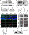
CX3CR1−/− mice suppressed microglial pyroptosis following ischemic stroke. (A and B) Representative immunoblotting imaging and quantitative analysis of GSDMD in the ischemic penumbra at 0, 1, 3, 5, 7, 14, 28, and 35 days after reperfusion, N = 6 per group. (C and D) Protein levels of GSDMD in CX3CR1+/− and CX3CR1−/− mice at 7 days after MCAO, N = 6 per group. (E and F): Representative immunofluorescence images of GSDMD co‐stain with Iba‐1 and GFAP in the ipsilateral hippocampus region, and quantitative analysis of GSDMD‐positive cells 7 days after reperfusion, N = 6 per group. (scale bar:100 μm and 20 μm). (G): Representative transmission electron microscopy images of membrane pores formed on microglia in the ipsilateral hippocampus region at 7 days after reperfusion, N = 3 in each group. (scale bar:2.0 μm and 1.0 μm). (H): The ELISA assay kit detected IL‐18 and IL‐1β production in the ipsilateral hippocampus. N = 6–8 per group. Data are shown as the mean ± SEM. *p < 0.05, **p < 0.01, ***p < 0.001 versus Sham+CX3CR1+/− group; # p < 0.05, ## p < 0.01, ### p < 0.001 versus MCAO+CX3CR1+/− group. Con: contralateral hippocampal regions; Ipsi: ipsilateral hippocampal regions.
FIGURE 5
FIGURE 5
CX3CR1−/− mice inhibited ischemia‐induced NLRP3 inflammasome activation and P65 signaling activation post‐ischemic stroke. (A and B): Representative immunoblotting imaging and quantitative analysis of NLRP, ASC, caspase‐1, IL‐18, and IL‐1β expression in the ipsilateral hippocampus region at 7 days after reperfusion, N = 6 per group. (C and D) Immunoblotting and quantitative analysis for p65 and p‐p65 in the ipsilateral hippocampus region 7 days after reperfusion, N = 6–8 per group. Data are shown as the mean ± SEM. *p < 0.05, **p < 0.01, ***p < 0.001 versus Sham+CX3CR1+/− group; # p < 0.05, ## p < 0.01, ### p < 0.001 versus MCAO+CX3CR1+/− group. Con: contralateral hippocampal regions; Ipsi: ipsilateral hippocampal regions.
FIGURE 6
FIGURE 6
CX3CR1−/− mice reduced brain damage and enhanced the rate of survival post‐ischemic stroke. (A and B) Representative images and quantification of Nissl staining at 7 days after reperfusion. N = 6 per group. (C and D) Representative images and quantification of [18F] DPA‐714 PET images at 7 days after reperfusion. N = 4 per group. (E and F) Representative images and the quantitative analysis of TTC staining at 3 days after reperfusion. N = 8 per group. All the data are shown as the mean ± SEM. *p < 0.05, **p < 0.01, ***p < 0.001 versus Sham+CX3CR1+/− group; # p < 0.05, ## p < 0.01, ### p < 0.001 versus MCAO+CX3CR1+/− group.

References

    1. Feigin VL, Nguyen G, Cercy K, et al. Global, regional, and country‐specific lifetime risks of stroke, 1990 and 2016. N Engl J Med. 2018;379:2429‐2437. - PMC - PubMed
    1. Campbell BCV, De Silva DA, Macleod MR, et al. Ischaemic stroke. Nat Rev Dis Primers. 2019;5:70. - PubMed
    1. Ferro JM, Caeiro L, Figueira ML. Neuropsychiatric sequelae of stroke. Nat Rev Neurol. 2016;12:269‐280. - PubMed
    1. Mijajlović MD, Pavlović A, Brainin M, et al. Post‐stroke dementia—a comprehensive review. BMC Med. 2017;15:11. - PMC - PubMed
    1. Rost NS, Brodtmann A, Pase MP, et al. Post‐stroke cognitive impairment and dementia. Circ Res. 2022;130:1252‐1271. - PubMed

Publication types