

Alterations in cortical volume and complexity in Parkinson's disease with depression
- PMID: 38421103
- PMCID: PMC10851315
- DOI: 10.1111/cns.14582
Alterations in cortical volume and complexity in Parkinson's disease with depression
Abstract
Aims: The aim of this study is to investigate differences in gray matter volume and cortical complexity between Parkinson's disease with depression (PDD) patients and Parkinson's disease without depression (PDND) patients.
Methods: A total of 41 PDND patients, 36 PDD patients, and 38 healthy controls (HC) were recruited and analyzed by Voxel-based morphometry (VBM) and surface-based morphometry (SBM). Differences in gray matter volume and cortical complexity were compared using the one-way analysis of variance (ANOVA) and correlated with the Hamilton Depression Scale-17 (HAMD-17) scores.
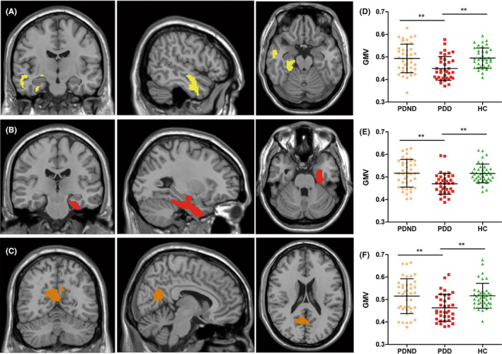
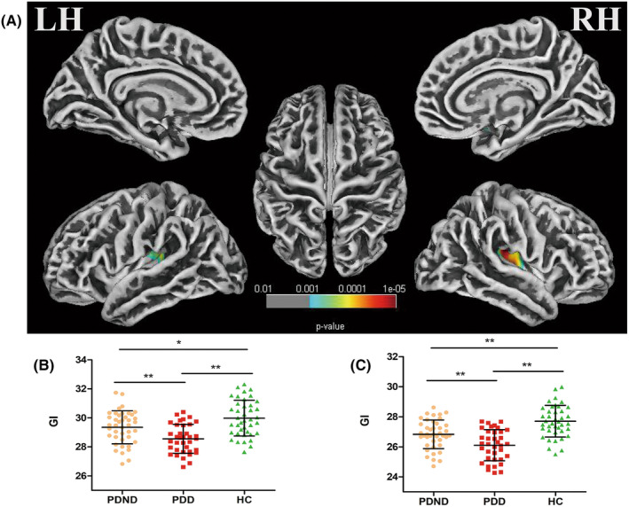
Results: PDD patients exhibited significant cortical atrophy in various regions, including bilateral medial parietal-occipital-temporal lobes, right dorsolateral temporal lobes, bilateral parahippocampal gyrus, and bilateral hippocampus, compared to HC and PDND groups. A negative correlation between the GMV of left precuneus and HAMD-17 scores in the PDD group tended to be significant (r = -0.318, p = 0.059). Decreased gyrification index was observed in the bilateral insular and dorsolateral temporal cortex. However, there were no significant differences found in fractal dimension and sulcal depth.
Conclusion: Our research shows extensive cortical structural changes in the insular cortex, parietal-occipital-temporal lobes, and hippocampal regions in PDD. This provides a morphological perspective for understanding the pathophysiological mechanism underlying depression in Parkinson's disease.
Keywords: Parkinson's disease; depression; structural magnetic resonance imaging; surface-based morphometry (SBM); voxel-based morphometry (VBM).
© 2023 The Authors. CNS Neuroscience & Therapeutics published by John Wiley & Sons Ltd.
Conflict of interest statement
The authors declare that the research was conducted in the absence of any commercial or financial relationships that could be construed as a potential conflict of interest.
Figures
Similar articles
-
Brain structural changes in diabetic retinopathy patients: a combined voxel-based morphometry and surface-based morphometry study.Brain Imaging Behav. 2024 Oct;18(5):1131-1143. doi: 10.1007/s11682-024-00905-7. Epub 2024 Aug 22. Brain Imaging Behav. 2024. PMID: 39172355
-
Voxel-based meta-analysis of gray matter volume reductions associated with cognitive impairment in Parkinson's disease.J Neurol. 2016 Jun;263(6):1178-87. doi: 10.1007/s00415-016-8122-3. Epub 2016 Apr 25. J Neurol. 2016. PMID: 27113603
-
Patterns of Grey Matter Atrophy at Different Stages of Parkinson's and Alzheimer's Diseases and Relation to Cognition.Brain Topogr. 2019 Jan;32(1):142-160. doi: 10.1007/s10548-018-0675-2. Epub 2018 Sep 11. Brain Topogr. 2019. PMID: 30206799
-
Voxelwise meta-analysis of gray matter anomalies in Parkinson variant of multiple system atrophy and Parkinson's disease using anatomic likelihood estimation.Neurosci Lett. 2015 Feb 5;587:79-86. doi: 10.1016/j.neulet.2014.12.007. Epub 2014 Dec 5. Neurosci Lett. 2015. PMID: 25484255
-
Gray matter atrophy in Parkinson's disease with dementia: evidence from meta-analysis of voxel-based morphometry studies.Neurol Sci. 2013 May;34(5):613-9. doi: 10.1007/s10072-012-1250-3. Epub 2012 Nov 27. Neurol Sci. 2013. PMID: 23184330 Review.
Cited by
-
Abnormal topological structure of structural covariance networks based on fractal dimension in noise induced hearing loss.Sci Rep. 2024 Nov 28;14(1):29644. doi: 10.1038/s41598-024-80731-5. Sci Rep. 2024. PMID: 39609512 Free PMC article.
References
Publication types
MeSH terms
Grants and funding
LinkOut - more resources
Full Text Sources
Medical
