

De novo variants predicting haploinsufficiency for DIP2C are associated with expressive speech delay
- PMID: 38421105
- PMCID: PMC11161320
- DOI: 10.1002/ajmg.a.63559
De novo variants predicting haploinsufficiency for DIP2C are associated with expressive speech delay
Abstract
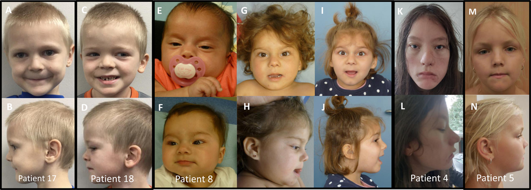
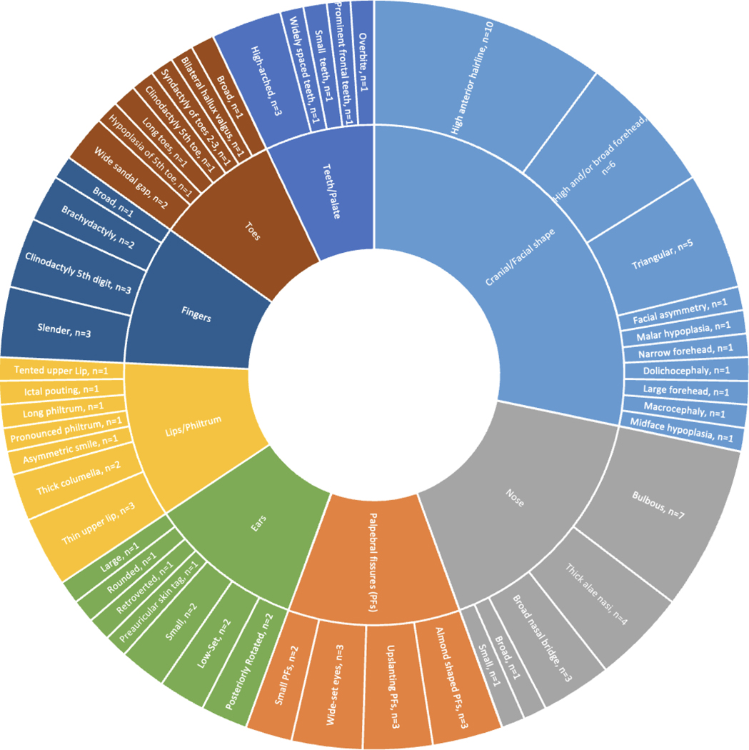
The disconnected (disco)-interacting protein 2 (DIP2) gene was first identified in D. melanogaster and contains a DNA methyltransferase-associated protein 1 (DMAP1) binding domain, Acyl-CoA synthetase domain and AMP-binding sites. DIP2 regulates axonal bifurcation of the mushroom body neurons in D. melanogaster and is required for axonal regeneration in the neurons of C. elegans. The DIP2 homologues in vertebrates, Disco-interacting protein 2 homolog A (DIP2A), Disco-interacting protein 2 homolog B (DIP2B), and Disco-interacting protein 2 homolog C (DIP2C), are highly conserved and expressed widely in the central nervous system. Although there is evidence that DIP2C plays a role in cognition, reports of pathogenic variants in these genes are rare and their significance is uncertain. We present 23 individuals with heterozygous DIP2C variants, all manifesting developmental delays that primarily affect expressive language and speech articulation. Eight patients had de novo variants predicting loss-of-function in the DIP2C gene, two patients had de novo missense variants, three had paternally inherited loss of function variants and six had maternally inherited loss-of-function variants, while inheritance was unknown for four variants. Four patients had cardiac defects (hypertrophic cardiomyopathy, atrial septal defects, and bicuspid aortic valve). Minor facial anomalies were inconsistent but included a high anterior hairline with a long forehead, broad nasal tip, and ear anomalies. Brainspan analysis showed elevated DIP2C expression in the human neocortex at 10-24 weeks after conception. With the cases presented herein, we provide phenotypic and genotypic data supporting the association between loss-of-function variants in DIP2C with a neurocognitive phenotype.
Keywords: DIP2; DIP2C; developmental delay; intellectual disability; speech articulation; speech delay.
© 2024 The Authors. American Journal of Medical Genetics Part A published by Wiley Periodicals LLC.
Figures


References
-
- Bowling KM, Thompson ML, Finnila CR, Hiatt SM, Latner DR, Amaral MD, Lawlor JMJ, East KM, Cochran ME, Greve V, Kelley WV, Gray DE, Felker SA, Meddaugh H, Cannon A, Luedecke A, Jackson KE, Hendon LG, Janani HM, Johnston M, Merin LA, Deans SL, Tuura C, Williams H, Laborde K, Neu MB, Patrick-Esteve J, Hurst ACE, Kandasamy J, Carlo W, Brothers KB, Kirmse BM, Savich R, Superneau D, Spedale SB, Knight SJ, Barsh GS, Korf BR, Cooper GM. Genome sequencing as a first-line diagnostic test for hospitalized infants. Genet Med. 2022. Apr;24(4):851–861. doi: 10.1016/j.gim.2021.11.020. Epub 2021 Nov 27. PMID: 34930662; PMCID: PMC8995345. - DOI - PMC - PubMed
-
- Cobben JM, Weiss MM, van Dijk FS, De Reuver R, de Kruiff C, Pondaag W, Hennekam RC, Yntema HG. A de novo mutation in ZMYND11, a candidate gene for 10p15.3 deletion syndrome, is associated with syndromic intellectual disability. Eur J Med Genet. 2014. Nov-Dec;57(11–12):636–8. doi: 10.1016/j.ejmg.2014.09.002. Epub 2014 Sep 30. PMID: 25281490. - DOI - PubMed
-
- Coe BP, Witherspoon K, Rosenfeld JA, van Bon BW, Vulto-van Silfhout AT, Bosco P, Friend KL, Baker C, Buono S, Vissers LE, Schuurs-Hoeijmakers JH, Hoischen A, Pfundt R, Krumm N, Carvill GL, Li D, Amaral D, Brown N, Lockhart PJ, Scheffer IE, Alberti A, Shaw M, Pettinato R, Tervo R, de Leeuw N, Reijnders MR, Torchia BS, Peeters H, O’Roak BJ, Fichera M, Hehir-Kwa JY, Shendure J, Mefford HC, Haan E, Gécz J, de Vries BB, Romano C, Eichler EE. Refining analyses of copy number variation identifies specific genes associated with developmental delay. Nat Genet. 2014. Oct;46(10):1063–71. doi: 10.1038/ng.3092. Epub 2014 Sep 14. PMID: 25217958; PMCID: PMC4177294. - DOI - PMC - PubMed
-
- DeScipio C, Spinner NB, Kaur M, Yaeger D, Conlin LK, Ambrosini A, Hu S, Shan S, Krantz ID, Riethman H. Fine-mapping subtelomeric deletions and duplications by comparative genomic hybridization in 42 individuals. Am J Med Genet A. 2008. Mar 15;146A(6):730–9. - PubMed
Publication types
MeSH terms
Substances
Grants and funding
LinkOut - more resources
Full Text Sources
Molecular Biology Databases
Research Materials
