. 2024 Apr 24;52(7):3823-3836.
doi: 10.1093/nar/gkae145.

The repertoire and structure of adhesion GPCR transcript variants assembled from publicly available deep-sequenced human samples

Affiliations

The repertoire and structure of adhesion GPCR transcript variants assembled from publicly available deep-sequenced human samples

Christina Katharina Kuhn et al. Nucleic Acids Res. .

Abstract

Alternative splicing and multiple transcription start and termination sites can produce a diverse repertoire of mRNA transcript variants from a given gene. While the full picture of the human transcriptome is still incomplete, publicly available RNA datasets have enabled the assembly of transcripts. Using publicly available deep sequencing data from 927 human samples across 48 tissues, we quantified known and new transcript variants, provide an interactive, browser-based application Splice-O-Mat and demonstrate its relevance using adhesion G protein-coupled receptors (aGPCRs) as an example. On average, 24 different transcript variants were detected for each of the 33 human aGPCR genes, and several dominant transcript variants were not yet annotated. Variable transcription starts and complex exon-intron structures encode a flexible protein domain architecture of the N- and C termini and the seven-transmembrane helix domain (7TMD). Notably, we discovered the first GPCR (ADGRG7/GPR128) with eight transmembrane helices. Both the N- and C terminus of this aGPCR were intracellularly oriented, anchoring the N terminus in the plasma membrane. Moreover, the assessment of tissue-specific transcript variants, also for other gene classes, in our application may change the evaluation of disease-causing mutations, as their position in different transcript variants may explain tissue-specific phenotypes.


Figures

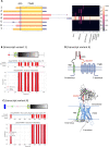
Graphical Abstract
Graphical Abstract
Figure 1.
Figure 1.
Tissue-specific transcript variants of ADGRD1/GPR133. (A) Transcript variants and ORFs of ADGRD1/GPR133 are depicted (orange: already annotated, red: newly identified). Protein domains depicted as yellow boxes. Heatmap shows relative expression of each transcript variant in % of the total transcript variant count per each tissue (Supplementary Table S5). (B) Contribution of 22 transcripts to overall ADGRD1 expression, each violin plot contains all 48 tissues. (C) Pearson correlation of absolute expression of transcript variants within ADGRD1, specifically transcript variants 2, 5, 8 and 13. The individual data points represent different tissues, 95% confidence intervals are shown. PTX, Pentraxin domain. GPS, G protein-coupled receptor proteolytic site. 7TMD, seven-transmembrane helix domain.
Figure 2.
Figure 2.
Structural variability of aGPCR isoforms resulting from different transcript variants. The structural variability of aGPCR isoforms are classified into isoforms with (A) NTF domain variability, (B) a variability in the C terminus, (C) variability in the 7TMD, (D) mRNA encoding for soluble N termini, (E) membrane-anchored N terminus and (F) CTF or substantially truncated N terminus. 7TMD, seven-transmembrane helix domain; CTF, C-terminal fragment; NTF, N-terminal fragment. The figure was created with BioRender.com.
Figure 3.
Figure 3.
Predicted transmembrane helix topology of ADGRG7/GPR128. (A) ADGRG7 has seven transcript variants, among which the novel transcript variant 4 (NSTRG.57932.4) showed highest expression in most tissues. The annotated transcript variants 1 and 2 (NM_001308362.1 and NM_032787.3) showed expression at significant levels in some tissues such as colon, small intestine, and liver. Heatmap shows relative expression of each transcript variant in % of the total transcript variant count per each tissue (Supplementary Table S5). Amino acid sequences of human ADGRG7 analyzed with SMART and DeepTMHMM predicted the GPS and 7TMD in all transcript variants and an additional transmembrane helix in transcript variants 2, 3 and 4 at the very N terminus. A signal peptide was only predicted in transcript variant 1 (B, red) and 6, however, not in transcript variant 4 (C). (D) In a hypothetical model of the ADGRG7 protein derived from transcript variant 4, the N- and C termini were located intracellularly. The three-dimensional model of the isoform derived from the long ADGRG7 transcript variant (Q96K78) generated by AlphaFold (AF-Q96K78-F1-model_v4.pdb) similarly revealed eight transmembrane helices, with the N terminus located intracellularly. The figure was partly created with BioRender.com.
Figure 4.
Figure 4.
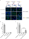
The N- and C terminus of ADGRG7/GPR128 is located intracellularly. COS-7 cells were transiently transfected with the double-tagged mouse ADGRG7/GPR128 and investigated in immunofluorescence (A) and ELISA (B, C). For immunofluorescence transfected cells were permeabilized and incubated with an antibody against the HA tag and an antibody against the C-terminal FLAG tag (as indicated for the constructs). The monoclonal antibodies were stained with a secondary goat anti-mouse IgG AlexaFluor488 nm-conjugated antibody (green). DAPI (blue) was used to visualize the nucleus and confocal images were taken with the BZ-X810 Fluorescence Microscope, Keyence with a 20x objective. Representative images are shown. The bar indicates 25 μm. Cell surface (B) and sandwich ELISA (C) tests were performed to determine the expression levels at the non-permeabilized plasma membrane and the solubilized full-length receptor, respectively. Shown is the mean ± SD of three independent assays performed in triplicate. The statistical significance was tested with a Student's t-test over the mean of biological replicates (n = 3). The figure was partly created with BioRender.com.
Figure 5.
Figure 5.
Projection of ADGRC1/CELSR1 mutations found in patients with neural tube defects. (Likely) pathogenic mutations from the Human Gene Mutation Database (HGMD®, https://www.hgmd.cf.ac.uk/ac/index.php) were projected onto the transcript variants. Depending on the position of the mutations, a subset of transcript variants may remain unaffected. Domain structure is given on top (generated by the SMART, see Materials and methods). Heatmap shows relative expression of each transcript variant in % of the total transcript variant count per each tissue (Supplementary Table S5).

References

    1. Alexander S.P.H., Christopoulos A., Davenport A.P., Kelly E., Mathie A., Peters J.A., Veale E.L., Armstrong J.F., Faccenda E., Harding S.D. et al. . THE CONCISE GUIDE TO PHARMACOLOGY 2019/20: g protein-coupled receptors. Br. J. Pharmacol. 2019; 176:S21–S141. - PMC - PubMed
    1. Markovic D., Challiss R.A.J. Alternative splicing of G protein-coupled receptors: physiology and pathophysiology. Cell. Mol. Life Sci. 2009; 66:3337–3352. - PMC - PubMed
    1. McMillan D.R., Kayes-Wandover K.M., Richardson J.A., White P.C. Very large G protein-coupled receptor-1, the largest known cell surface protein, is highly expressed in the developing central nervous system. J. Biol. Chem. 2002; 277:785–792. - PubMed
    1. Knierim A.B., Röthe J., Çakir M.V., Lede V., Wilde C., Liebscher I., Thor D., Schöneberg T. Genetic basis of functional variability in adhesion G protein-coupled receptors. Sci. Rep. 2019; 9:11036. - PMC - PubMed
    1. Liebscher I., Schön J., Petersen S.C., Fischer L., Auerbach N., Demberg L.M., Mogha A., Cöster M., Simon K.-U., Rothemund S. et al. . A tethered agonist within the ectodomain activates the adhesion G protein-coupled receptors GPR126 and GPR133. Cell Rep. 2014; 9:2018–2026. - PMC - PubMed

Publication types

MeSH terms

Substances