. 2024 Mar 15;5(1):102925.
doi: 10.1016/j.xpro.2024.102925. Epub 2024 Feb 28.

Protocol for differentiation of functional macrophages from human induced pluripotent stem cells

Affiliations

Protocol for differentiation of functional macrophages from human induced pluripotent stem cells

Suji Jeong et al. STAR Protoc. .

Abstract

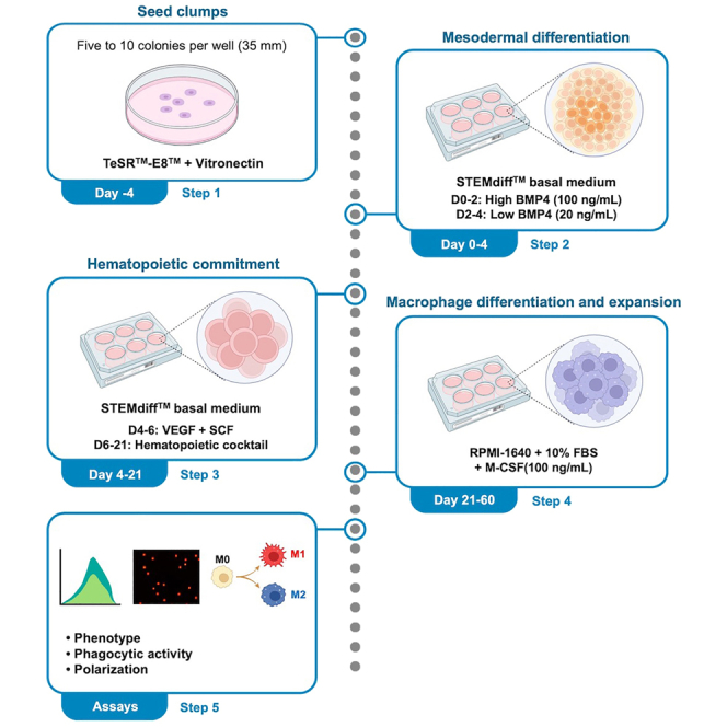
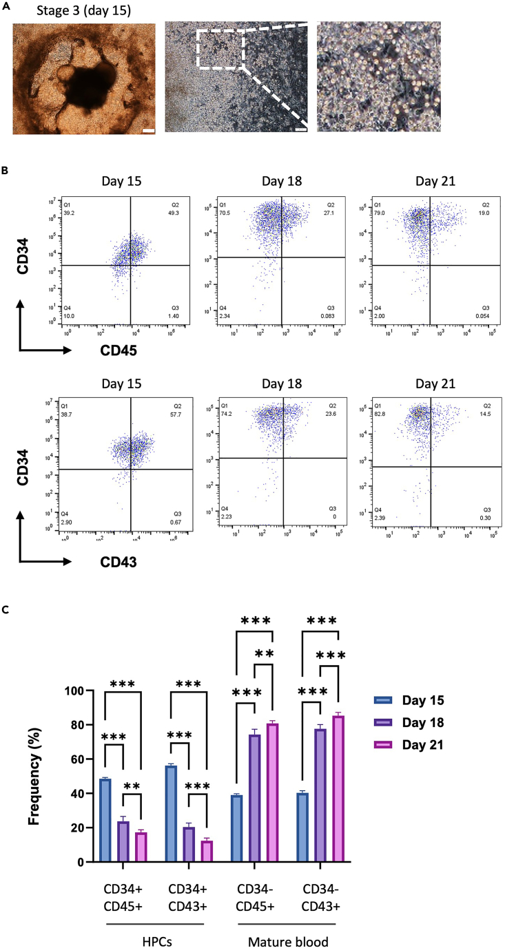
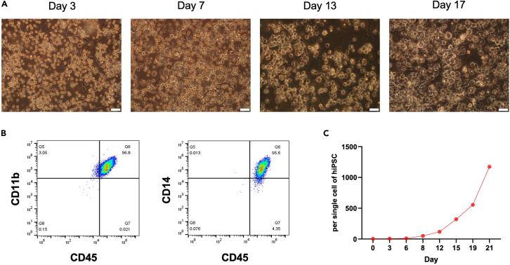
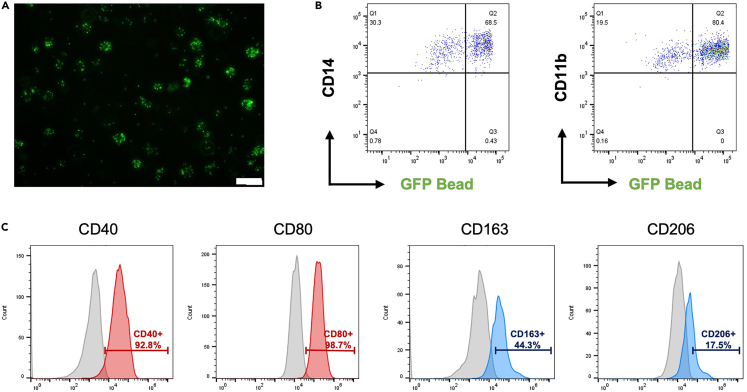
Human induced pluripotent stem cell (hiPSC)-derived macrophages provide a valuable tool for disease modeling and drug discovery. Here, we present a protocol to generate functional macrophages from hiPSCs using a feeder-free hematopoietic differentiation technique. We describe steps for preparing hiPSCs, mesodermal differentiation, hematopoietic commitment, and macrophage differentiation and expansion. We then detail assays to characterize their phenotype, polarization, and phagocytic functions. The functional macrophages generated here could be used to generate organoids for disease modeling and drug discovery studies. For complete details on the use and execution of this protocol, please refer to Jeong et al.1 and Heo et al.2.

Keywords: Cell Biology; Cell Differentiation; Cell culture; Cell isolation; Stem Cells.


Conflict of interest statement

Declaration of interests The mesodermal induction with high dose of BMP4 and subsequent hematopoietic and macrophage differentiation are protected by published patents (KOR/10-2106710), and these intellectual property rights belong to KW-Bio Co., Ltd.

Figures

None
Graphical abstract
Figure 1
Figure 1
Example images of healthy and differentiated colonies that are maintained in culture prior to the onset of differentiation Healthy colonies are densely composed of homogeneous single cells and appear as a single layer (left image). In differentiated colonies, a cluster of brightly glowing cells is produced from the center of the colony (right image, white arrowhead). These colonies will increase in size over time, and if there are differentiated colonies present, they will be less efficient at differentiating the desired hematopoietic progenitor cells. Scale bars are 200 μm.
Figure 2
Figure 2
Schematic overview of all steps of the protocol Schematic overview of the four-step protocol for the induction of hematopoietic cells and macrophages from hiPSCs.
Figure 3
Figure 3
Effect of BMP4 treatment concentration on early mesoderm differentiation (A) Representative bright-field images of colonies in which the early mesoderm was induced with low (20 ng/mL) and high (100 ng/mL) concentrations of BMP4 in Stage 1. Scale bars are 200 μm. (B) Differentiation days 0–2. The differences in mesoderm differentiation markers (T and MIXL1) between low (20 ng/mL) and high (100 ng/mL) doses of BMP4 were confirmed by quantitative reverse transcriptase polymerase chain reaction (qRT–PCR). The bars indicate the mean ± SD, n = 3, ∗p < 0.05, ∗∗p < 0.01, ∗∗∗p < 0.001. UN, undifferentiated hiPSCs. (C) Representative bright-field images of colonies during the hemogenic specification differentiation of hiPSCs. Scale bar is 200 μm.
Figure 4
Figure 4
Changes in the morphology and phenotype of HPCs during the hematopoietic commitment phase (A) Representative bright-field images of colonies (upper) and floating HPCs (lower) during the hematopoietic commitment phase of hiPSCs. Scale bars are 200 μm (upper) and 100 μm (lower). (B) Representative FACS dot plots of CD34, CD43, and CD45 expression in generated HPCs. (C) The generated HPCs were analyzed by flow cytometry for the phenotypes of primitive hematopoietic progenitors (CD34+CD45+ or CD34+CD43+) and mature blood cells (CD34-CD45+ or CD34-CD43+) during days 15–21. The bars indicate the mean ± SD, ∗∗p < 0.01, ∗∗∗p < 0.001.
Figure 5
Figure 5
Morphological changes in HPCs following macrophage differentiation (A) Representative bright-field images of HPC from days 3–17 as they differentiate into macrophages. Scale bars are 50 μm. (B) Macrophage phenotype markers (CD45+CD11b+, CD45+CD14+) were measured by flow cytometry, and data were analyzed using FlowJo software (Tree Star). (C) The graph shows the number of hiPSC-derived macrophages over the three weeks. The proliferation rate per 1 well was calculated by counting the number of macrophage cells.
Figure 6
Figure 6
Analysis of the phagocytosis activity and polarization of macrophages (A) Representative fluorescence image of the macrophage uptake of GFP-labeled carboxylate-modified polystyrene latex beads. Scale bar is 50 μm. (B) Macrophage phagocytosis was analyzed by flow cytometry using GFP-labeled carboxylate-modified polystyrene latex beads. Frequencies for macrophage markers (CD14 and CD11b) and GFP-labeled carboxylate-modified polystyrene latex beads were measured by flow cytometry. (C) The phenotypes of M1 (CD40, CD80) and M2 (CD163, CD206) polarized macrophages were measured by flow cytometry. The data were analyzed using FlowJo software (Tree Star).

References

    1. Jeong S., An B., Kim J.H., Han H.W., Kim J.H., Heo H.R., Ha K.S., Han E.T., Park W.S., Hong S.H. BMP4 and perivascular cells promote hematopoietic differentiation of human pluripotent stem cells in a differentiation stage-specific manner. Exp. Mol. Med. 2020;52:56–65. doi: 10.1038/s12276-019-0357-5. - DOI - PMC - PubMed
    1. Heo H.R., Hong S.H. Generation of macrophage containing alveolar organoids derived from human pluripotent stem cells for pulmonary fibrosis modeling and drug efficacy testing. Cell Biosci. 2021;11:216. doi: 10.1186/s13578-021-00721-2. - DOI - PMC - PubMed
    1. Heo H.R., Song H., Kim H.R., Lee J.E., Chung Y.G., Kim W.J., Yang S.R., Kim K.S., Chun T., Lee D.R., Hong S.H. Reprogramming mechanisms influence the maturation of hematopoietic progenitors from human pluripotent stem cells. Cell Death Dis. 2018;9:1090. doi: 10.1038/s41419-018-1124-6. - DOI - PMC - PubMed
    1. Kim J.H., Jo H.Y., Ha H.Y., Kim Y.O. Korea National Stem Cell Bank. Stem Cell Res. 2021;53 doi: 10.1016/j.scr.2021.102270. - DOI - PubMed
    1. Lim J.J., Kim H.J., Rhie B.H., Lee M.R., Choi M.J., Hong S.H., Kim K.S. Maintenance of hPSCs under Xeno-Free and Chemically Defined Culture Conditions. Int. J. Stem Cells. 2019;12:484–496. doi: 10.15283/ijsc19090. - DOI - PMC - PubMed

Publication types

LinkOut - more resources