. 2024 Mar 5;121(10):e2317147121.
doi: 10.1073/pnas.2317147121. Epub 2024 Feb 29.

Pathogenic GATA2 genetic variants utilize an obligate enhancer mechanism to distort a multilineage differentiation program

Affiliations

Pathogenic GATA2 genetic variants utilize an obligate enhancer mechanism to distort a multilineage differentiation program

Koichi R Katsumura et al. Proc Natl Acad Sci U S A. .

Abstract

Mutations in genes encoding transcription factors inactivate or generate ectopic activities to instigate pathogenesis. By disrupting hematopoietic stem/progenitor cells, GATA2 germline variants create a bone marrow failure and leukemia predisposition, GATA2 deficiency syndrome, yet mechanisms underlying the complex phenotypic constellation are unresolved. We used a GATA2-deficient progenitor rescue system to analyze how genetic variation influences GATA2 functions. Pathogenic variants impaired, without abrogating, GATA2-dependent transcriptional regulation. Variants promoted eosinophil and repressed monocytic differentiation without regulating mast cell and erythroid differentiation. While GATA2 and T354M required the DNA-binding C-terminal zinc finger, T354M disproportionately required the N-terminal finger and N terminus. GATA2 and T354M activated a CCAAT/Enhancer Binding Protein-ε (C/EBPε) enhancer, creating a feedforward loop operating with the T-cell Acute Lymphocyte Leukemia-1 (TAL1) transcription factor. Elevating C/EBPε partially normalized hematopoietic defects of GATA2-deficient progenitors. Thus, pathogenic germline variation discriminatively spares or compromises transcription factor attributes, and retaining an obligate enhancer mechanism distorts a multilineage differentiation program.

Keywords: GATA; GATA2; differentiation; hematopoiesis; transcription.


Conflict of interest statement

Competing interests statement:The authors declare no competing interest.

Figures
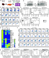
Fig. 1.
Fig. 1.
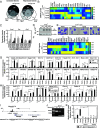
GATA2 disease variants are defective in regulating differentiation and transcription in a genetic rescue system. (A) Schematic representation of the genetic rescue system. (B) Left: Representative western blot analysis of −77−/− fetal liver cells expressing HA-GATA2 or variants with anti-HA antibody. Right: Representative western blot analysis of −77+/+ and −77−/− fetal liver cells expressing HA-GATA2 or variants with anti-GATA2 antibody. (C) Flow cytometric analysis of GFP+ cells stained for monocytic cells (CD11b+CD115+), granulocytic cells (CD11b+CD115), macrophage (F4/80+CD11b+), mast cells (FcεR1α+Kit+), and eosinophils (CD11b+SiglecF+). (D) Quantitation of flow cytometry data (n = 5). (E) Heatmap showing the result of RT-qPCR analysis of erythroid, mast cell, and eosinophil genes (n = 5). (F) Flow cytometric analysis of GFP+ cells stained for monocytic cells (CD11b+CD115+), granulocytic cells (CD11b+CD115), macrophages (F4/80+CD11b+), mast cells (FcεR1α+Kit+), and eosinophils (CD11b+SiglecF+). (G) Quantitation of flow cytometry data (n = 4). * < 0.05, ** < 0.01, and *** < 0.001.
Fig. 2.
Fig. 2.
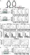
Context-dependent T354M occupancy of chromatin sites and transcriptional regulation. (A) Venn diagram depicting overlap between GATA2- and disease variant-regulated genes. (B) Heatmap depicting RT-qPCR analysis of GATA2-regulated genes (n = 5). (C) RT-qPCR analysis of GATA2-regulated mRNAs and primary transcripts in GATA2- or T354M-expressing cells (n = 5). (D) Representative western blot analysis of hi-77−/− cells expressing HA-GATA2 or variants with anti-HA antibody. (E) Representative image of Giemsa-stained cells. Cells were cultured for 3 d. (F) Heatmap depicting RT-qPCR analysis of GATA2-regulated mRNAs identified with GATA2- or disease variant-expressing hi-77−/− cells (n = 6). (G) Quantitative ChIP analysis at GATA2-occupied sites in target genes with anti-HA antibody (n = 5). (H) Quantitative ChIP analysis at GATA2-occupied sites in target genes with anti-GATA2 antibody (n = 4). (I) CUT&RUN profile of GATA2 at Ms4a3 locus (GSE171384). (J) PCR-genotyping assay for −77−/−Ms4a3+27.6−/− allele. (K) RT-qPCR analysis of Ms4a3 mRNA levels (n = 4). * < 0.05, ** < 0.01, and *** < 0.001.
Fig. 3.
Fig. 3.
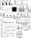
Zinc finger requirements for T354M-mediated transcriptional regulation. (A) Schematic representation of zinc finger structures. (B) Left, Representative western blot analysis of HA-GATA2 or variants using anti-HA antibody. Right, RT-qPCR analysis of GATA2-regulated mRNAs in cells expressing GATA2 or variants (n = 5). (C) Left, Representative western blot analysis of HA-GATA2 or variants using anti-HA antibody. Right, RT-qPCR analysis of GATA2-regulated mRNAs in cells expressing GATA2 or variants (n = 5). (D) RT-qPCR analysis of T354M-regulated mRNAs in cells expressing GATA2 or variants (n = 5). (E) Quantitative ChIP analysis at GATA-occupied sites in target genes with anti-HA antibody (n = 3). (F) RT-qPCR analysis of Irf8 mRNA in cells expressing GATA2 or variants (n = 5). (G) Left, Representative western blot analysis of HA-GATA2 or variants using anti-HA antibody. Right, RT-qPCR analysis of GATA2-regulated mRNAs in GATA2- or variant-expressing cells (n = 4). * < 0.05, ** < 0.01, and *** < 0.001.
Fig. 4.
Fig. 4.
Genetic ablation of TAL1 reveals its context-dependent requirement for GATA2 and T354M function. (A) Quantitative ChIP analysis with anti-TAL1 antibody at GATA2-occupied sites in target genes (n = 4). (B) Quantitative ChIP analysis with anti-LDB1 antibody at GATA2-occupied sites in target genes (n = 4). (C) PCR-based genotyping assay for −77−/−Tal1−/− allele. (D) Top, Representative western blot analysis of TAL1. Bottom, RT-qPCR analysis of Tal1 mRNA level in −77−/− and −77−/−Tal1−/− cells. (E) RT-qPCR analysis of GATA2 target gene mRNAs in cells expressing GATA2 or variants (n = 5). (F) Quantitative ChIP analysis at GATA2-occupied sites in target genes with anti-HA antibody (n = 3). (G) Model of collaborative transcriptional regulation by GATA2/T354M and TAL1. * < 0.05, ** < 0.01, and *** < 0.001.
Fig. 5.
Fig. 5.
Genetic ablation of a Cebpe enhancer reveals the vital importance of C/EBPε for context-dependent GATA2 and T354M function. (A) Left, Comparison of protein levels [normalized using MaxLFQ from the prior analysis (43)] in CMP/GMP. Center, mRNA levels (TPM) in Lin progenitors calculated using RSEM (68). Right, mRNA levels (TPM) in hi-77+/+ or hi-77−/− cell lines. (B) RT-qPCR analysis of Cebpe mRNA in cells expressing GATA2 or variants (n = 5). (C) ChIP-Seq profile of transcription factors at Cebpe locus in hi-77+/+ cells mined from dataset GEO:GSE224904. (D) PCR-genotyping assay for −77−/−Cebpe+6−/− allele. (E) Left, Representative western blot analysis of C/EBPε. Right: Quantification of C/EBPε protein in hi-77−/− cells expressing HA-GATA2 or variants (n = 4). (F) RT-qPCR analysis of GATA2 target gene mRNAs in cells expressing GATA2 or variants (n = 4). (G) ChIP-Seq profile of transcription factors at Prg2/3, Epx, Ms4a2/3, Csf2rb, and Anxa1 in mouse myelocytes mined from dataset GEO:GSE73844. (H) RT-qPCR analysis of GATA2 target gene mRNAs in hi-77+/+ cells and hi-77+/+Cebpe+6−/− cells (n = 6). (I) Representative western blot analysis of HA-C/EBPε by using anti-HA antibody. Right, RT-qPCR analysis of C/EBPε target gene mRNAs (n = 5). (J) Top, Flow cytometric analysis of GFP+ cells stained with CD11b and CD115. Bottom, Quantitation of flow cytometry data (n = 5). * < 0.05, ** < 0.01, and *** < 0.001.
Fig. 6.
Fig. 6.
Pathogenic GATA2 genetic variants utilize an obligate enhancer-dependent feedforward mechanism to distort a multilineage differentiation program. GATA2 and T354M utilize C/EBPε and TAL1 to regulate a target gene cohort and multilineage differentiation. The fragmented activity of T354M generates a lineage-distorted differentiation program involving increased eosinophil, but not erythroid or mast cell, differentiation. As other GATA2 pathogenic variants share this attribute, this may represent a hallmark of GATA2 pathogenic variants. Considering the involvement of C/EBPε as a suppressor of MLL-rearrangement AML (70), and GATA2 as a suppressor of MDS/AML, the GATA2-C/EBPε paradigm links two vital systems that ensure the normal development and function of the hematopoietic system.

Similar articles

Cited by

References

    1. Weinreb J. T., Gupta V., Sharvit E., Weil R., Bowman T. V., Ddx41 inhibition of DNA damage signaling permits erythroid progenitor expansion in zebrafish. Haematologica 107, 644–654 (2022). - PMC - PubMed
    1. Zhang Z., et al. , The helicase DDX41 senses intracellular DNA mediated by the adaptor STING in dendritic cells. Nat. Immunol. 12, 959–965 (2011). - PMC - PubMed
    1. Parvatiyar K., et al. , The helicase DDX41 recognizes the bacterial secondary messengers cyclic di-GMP and cyclic di-AMP to activate a type I interferon immune response. Nat. Immunol. 13, 1155–1161 (2012). - PMC - PubMed
    1. Tsai F. Y., et al. , An early haematopoietic defect in mice lacking the transcription factor GATA-2. Nature 371, 221–226 (1994). - PubMed
    1. Polprasert C., et al. , Inherited and somatic defects in DDX41 in myeloid neoplasms. Cancer Cell 27, 658–670 (2015). - PMC - PubMed

Substances

LinkOut - more resources