Mechanism of branching morphogenesis inspired by diatom silica formation
- PMID: 38422023
- PMCID: PMC10927588
- DOI: 10.1073/pnas.2309518121
Mechanism of branching morphogenesis inspired by diatom silica formation
Abstract
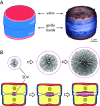
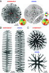
The silica-based cell walls of diatoms are prime examples of genetically controlled, species-specific mineral architectures. The physical principles underlying morphogenesis of their hierarchically structured silica patterns are not understood, yet such insight could indicate novel routes toward synthesizing functional inorganic materials. Recent advances in imaging nascent diatom silica allow rationalizing possible mechanisms of their pattern formation. Here, we combine theory and experiments on the model diatom Thalassiosira pseudonana to put forward a minimal model of branched rib patterns-a fundamental feature of the silica cell wall. We quantitatively recapitulate the time course of rib pattern morphogenesis by accounting for silica biochemistry with autocatalytic formation of diffusible silica precursors followed by conversion into solid silica. We propose that silica deposition releases an inhibitor that slows down up-stream precursor conversion, thereby implementing a self-replicating reaction-diffusion system different from a classical Turing mechanism. The proposed mechanism highlights the role of geometrical cues for guided self-organization, rationalizing the instructive role for the single initial pattern seed known as the primary silicification site. The mechanism of branching morphogenesis that we characterize here is possibly generic and may apply also in other biological systems.
Keywords: biomineralization; branching morphogenesis; diatom; silica patterning.
Conflict of interest statement
Competing interests statement:The authors declare no competing interest.
Figures


Similar articles
-
Reconstituting the formation of hierarchically porous silica patterns using diatom biomolecules.J Struct Biol. 2018 Oct;204(1):64-74. doi: 10.1016/j.jsb.2018.07.005. Epub 2018 Jul 29. J Struct Biol. 2018. PMID: 30009877
-
Nanopatterned protein microrings from a diatom that direct silica morphogenesis.Proc Natl Acad Sci U S A. 2011 Feb 22;108(8):3175-80. doi: 10.1073/pnas.1012842108. Epub 2011 Feb 7. Proc Natl Acad Sci U S A. 2011. PMID: 21300899 Free PMC article.
-
Shedding light on silica biomineralization by comparative analysis of the silica-associated proteomes from three diatom species.Plant J. 2022 Jun;110(6):1700-1716. doi: 10.1111/tpj.15765. Epub 2022 Apr 26. Plant J. 2022. PMID: 35403318
-
Silica biomineralization in diatoms: the model organism Thalassiosira pseudonana.Chembiochem. 2008 May 23;9(8):1187-94. doi: 10.1002/cbic.200700764. Chembiochem. 2008. PMID: 18381716 Review.
-
Prescribing diatom morphology: toward genetic engineering of biological nanomaterials.Curr Opin Chem Biol. 2007 Dec;11(6):662-9. doi: 10.1016/j.cbpa.2007.10.009. Epub 2007 Nov 26. Curr Opin Chem Biol. 2007. PMID: 17991447 Review.
Cited by
-
Reminiscent of the pre-diatom? A hitherto undescribed scaly bolidophyte Lepidoparma frigida gen. et sp. nov. in a new order Lepidoparmales based on morphology, phylogeny, and ecology.J Phycol. 2025 Aug;61(4):757-776. doi: 10.1111/jpy.70043. Epub 2025 Jun 19. J Phycol. 2025. PMID: 40536273 Free PMC article.
-
Intracellular morphogenesis of diatom silica is guided by local variations in membrane curvature.Nat Commun. 2024 Sep 10;15(1):7888. doi: 10.1038/s41467-024-52211-x. Nat Commun. 2024. PMID: 39251596 Free PMC article.
-
Hexagonal Patterns in Diatom Silica Form via a Directional Two-Step Process.Adv Sci (Weinh). 2024 Nov;11(41):e2402492. doi: 10.1002/advs.202402492. Epub 2024 Sep 6. Adv Sci (Weinh). 2024. PMID: 39239810 Free PMC article.
References
-
- Falkowski P. G., Barber R. T., Smetacek V., Biogeochemical controls and feedbacks on ocean primary production. Science 281, 200–206 (1998). - PubMed
-
- Field C. B., Behrenfeld M. J., Randerson J. T., Falkowski P., Primary production of the biosphere: Integrating terrestrial and oceanic components. Science 281, 237–240 (1998). - PubMed
-
- Hamm C. E., et al. , Architecture and material properties of diatom shells provide effective mechanical protection. Nature 421, 841–843 (2003). - PubMed
MeSH terms
Substances
Grants and funding
LinkOut - more resources
Full Text Sources

