. 2024 May 1;42(13):1477-1487.
doi: 10.1200/JCO.23.00295. Epub 2024 Feb 29.

Comprehensive Inherited Risk Estimation for Risk-Based Breast Cancer Screening in Women

Collaborators, Affiliations

Comprehensive Inherited Risk Estimation for Risk-Based Breast Cancer Screening in Women

Nina Mars et al. J Clin Oncol. .

Abstract

Purpose: Family history (FH) and pathogenic variants (PVs) are used for guiding risk surveillance in selected high-risk women but little is known about their impact for breast cancer screening on population level. In addition, polygenic risk scores (PRSs) have been shown to efficiently stratify breast cancer risk through combining information about common genetic factors into one measure.

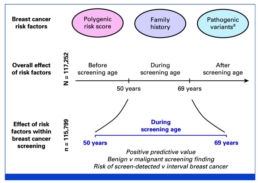
Methods: In longitudinal real-life data, we evaluate PRS, FH, and PVs for stratified screening. Using FinnGen (N = 117,252), linked to the Mass Screening Registry for breast cancer (1992-2019; nationwide organized biennial screening for age 50-69 years), we assessed the screening performance of a breast cancer PRS and compared its performance with FH of breast cancer and PVs in moderate- (CHEK2)- to high-risk (PALB2) susceptibility genes.

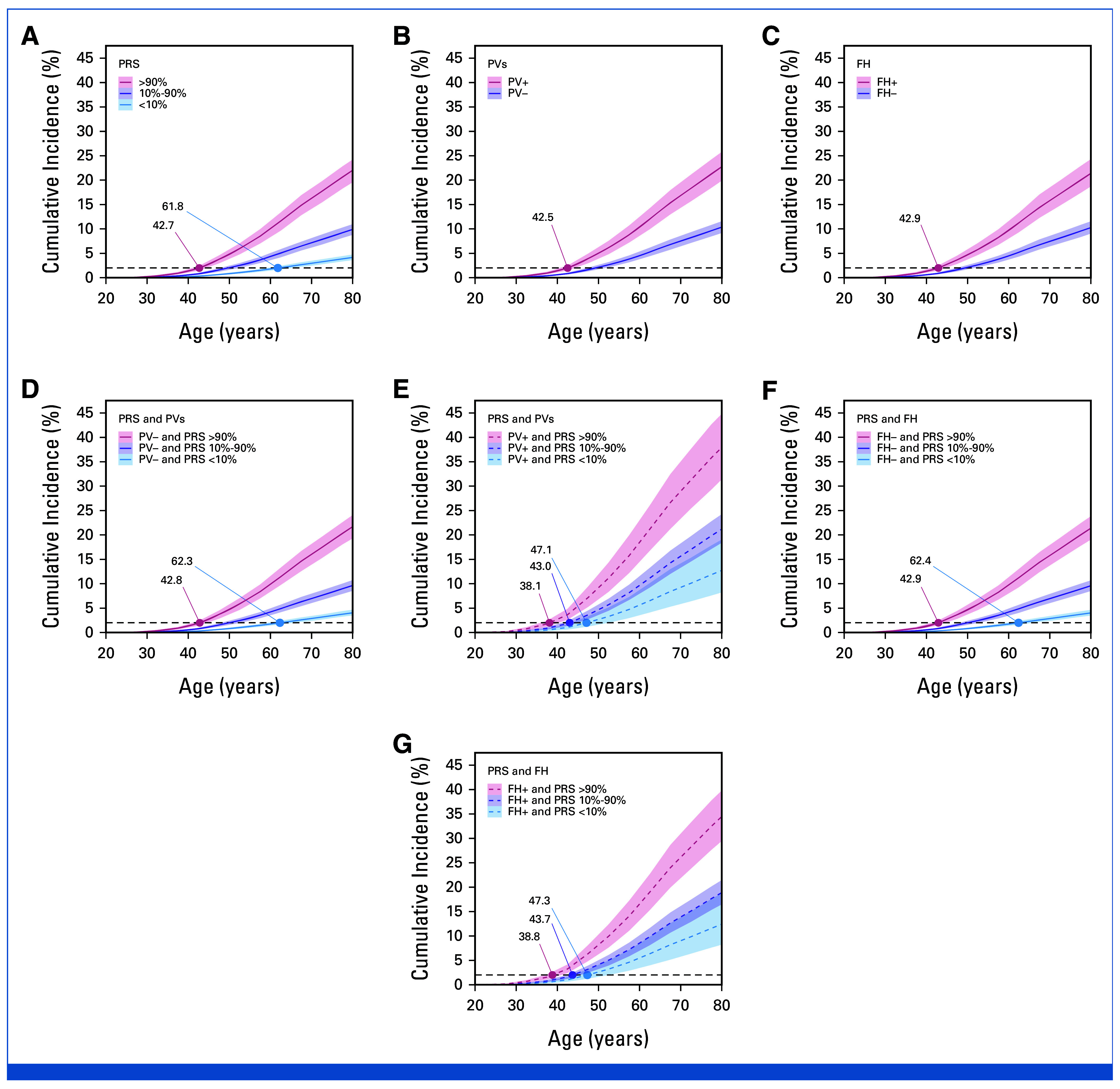
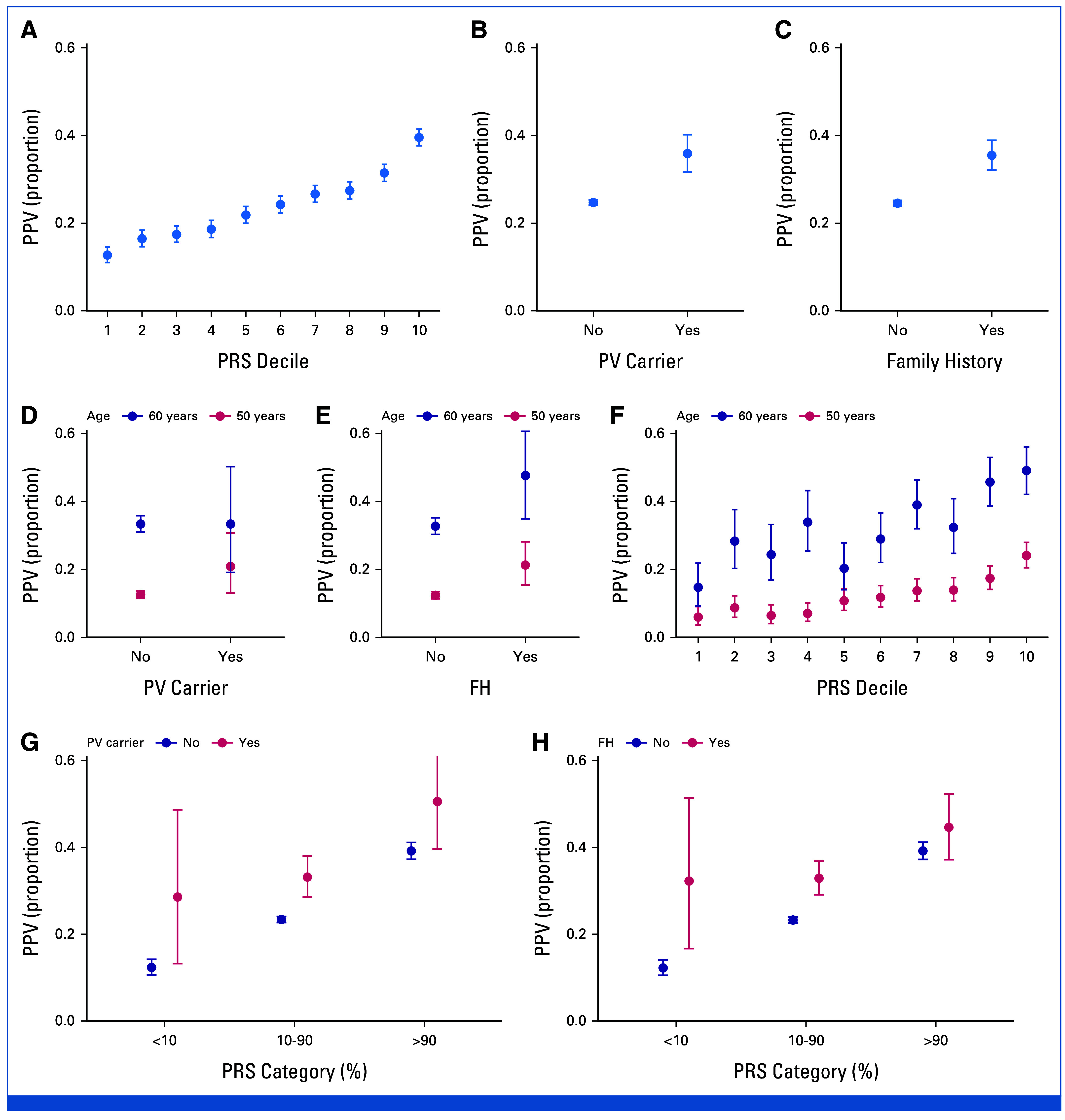
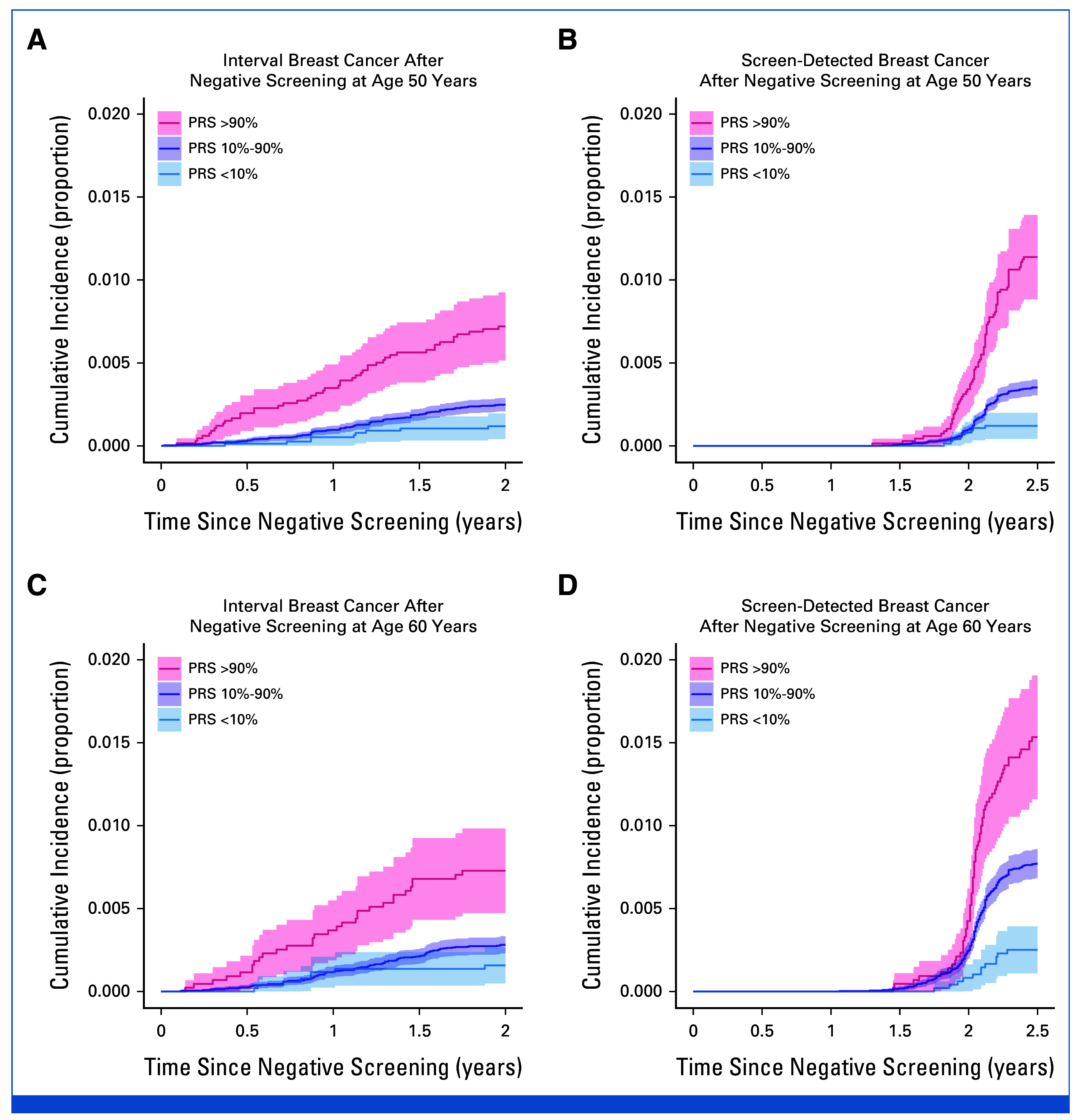
Results: Effect sizes for FH, PVs, and high PRS (>90th percentile) were comparable in screening-aged women, with similar implications for shifting age at screening onset. A high PRS identified women more likely to be diagnosed with breast cancer after a positive screening finding (positive predictive value [PPV], 39.5% [95% CI, 37.6 to 41.5]). Combinations of risk factors increased the PPVs up to 45% to 50%. A high PRS conferred an elevated risk of interval breast cancer (hazard ratio [HR], 2.78 [95% CI, 2.00 to 3.86] at age 50 years; HR, 2.48 [95% CI, 1.67 to 3.70] at age 60 years), and women with a low PRS (<10th percentile) had a low risk for both interval- and screen-detected breast cancers.

Conclusion: Using real-life screening data, this study demonstrates the effectiveness of a breast cancer PRS for risk stratification, alone and combined with FH and PVs. Further research is required to evaluate their impact in a prospective risk-stratified screening program, including cost-effectiveness.


Conflict of interest statement

The following represents disclosure information provided by authors of this manuscript. All relationships are considered compensated unless otherwise noted. Relationships are self-held unless noted. I = Immediate Family Member, Inst = My Institution. Relationships may not relate to the subject matter of this manuscript. For more information about ASCO's conflict of interest policy, please refer to www.asco.org/rwc or ascopubs.org/jco/authors/author-center.

Open Payments is a public database containing information reported by companies about payments made to US-licensed physicians (Open Payments).

Figures

FIG 1.
FIG 1.
Study overview. aMost frequent susceptibility genes in Finland (CHEK2 and PALB2).
FIG 2.
FIG 2.
Lifetime risk of breast cancer for categories of (A) PRS, by carrier status for (B) PV, (C) FH status, (D and E) the combination of PRS and PVs, and (F and G) the combination of PRS and FH. The numbers indicate when each category reaches a 2% cumulative incidence, aligning with the population's average at the start of organized screening at age 50 years. The cumulative incidences by PRS decile are provided in the Data Supplement (Fig S1). The estimates are calibrated to the general population, with the estimates accounting for competing risks of death from other causes than breast cancer (see the Data Supplement for details). FH, family history; PRS, polygenic risk score; PV, pathogenic variant.
FIG 3.
FIG 3.
Observed PPV (proportion of screen-detected cancers among women with a screening finding defined as women called back for complementary follow-up examination for an abnormal screening mammogram; see Methods for details) with different risk factors. The PPV (A) by PRS decile, (B) by PV carrier status, and (C) by FH status, with corresponding PPVs for two screening ages, (D-F) 50 and 60 years. (D-F) Also highlight the impact of age on PPV, because of differences in baseline risk of breast cancer, which increases with age. (G and H) The PPV by combinations of (G) PRS and PV carrier and (H) PRS and FH status, with PRS divided into three categories. PPVs at all screening ages (biennially from age 50 years to 69 years) are provided in the Data Supplement (Fig S2), and the detailed numbers for the Figure are provided in the Data Supplement (Table S1). Error bars show the 95% CIs. PVs: CHEK2 c.1100delC, CHEK2 c.319+2T>A, PALB2 c.1592delT, analyzed jointly for power, and heterozygotes were considered jointly with homozygotes. FH, family history; PPV, positive predictive value; PRS, polygenic risk score; PV, pathogenic variant.
FIG 4.
FIG 4.
Proportion of individuals with risk factors by type of most severe screening finding. Women with no finding have never had a positive screening finding. (A) Prevalence of women in different PRS deciles by screening finding. Each PRS decile contains by definition 10% of individuals (dotted line), whereby proportions over 10% indicate enrichment of the histologic finding. For instance, of women with bilateral breast cancer, 35.2% (95% CI, 27.4 to 43.5) had a PRS in the top decile. (B) Prevalence of each risk factor (PRS category, PV carrier, and family history) in women by screening finding. The PRS >90% contains the same information as the top deciles for each screening finding in (A). In addition to the final breast cancer diagnosis available in the Finnish Cancer Registry, information on histology from screening biopsies is available within the Mass Screening Registry, on the basis of ICD-O-3 morphology codes. The screening findings were divided into the following categories, with each woman classified on the basis of her most severe screening finding: (1) no finding (ie, no suspicion of malignancy, N = 110,185), (2) benign (nonmalignant) breast lesion (N = 758), (3) in situ lesion (N = 613), (4) unilateral invasive breast cancer (N = 4,098), and (5) bilateral breast cancer (N = 145; in situ or invasive lesions simultaneously in both breasts). Because of a small case count, the prevalence of PRS deciles one to three for bilateral breast cancer was calculated by pooling their prevalence and dividing it by three. Error bars show the 95% CIs. The P value for trend was calculated by randomly sampling 1,000 individuals in screening finding categories that had over 1,000 individuals (the categories were no finding and unilateral invasive breast cancer). PVs: CHEK2 c.1100delC, CHEK2 c.319+2T>A, PALB2 c.1592delT, analyzed jointly for power, and heterozygotes were considered jointly with homozygotes. ICD-O, International Classification of Diseases for Oncology; PRS, polygenic risk score; PV, pathogenic variant.
FIG 5.
FIG 5.
Survival curves showing cumulative incidence of breast cancer for interval and screen-detected cancers after a negative screen at age (A and B) 50 years and (C and D) 60 years by PRS category. Survival curves for a broader set of screening ages are shown in the Data Supplement (Fig S3). Cumulative incidence represents the proportion of individuals diagnosed by each time point shown on the x-axis. PRS, polygenic risk score.

References

    1. Iwamoto Y, Kaucher S, Lorenz E, et al. Development of breast cancer mortality considering the implementation of mammography screening programs—A comparison of western European countries. BMC Public Health. 2019;19:823. - PMC - PubMed
    1. Zielonke N, Gini A, Jansen EEL, et al. Evidence for reducing cancer-specific mortality due to screening for breast cancer in Europe: A systematic review. Eur J Cancer. 2020;127:191–206. - PubMed
    1. Clift AK, Dodwell D, Lord S, et al. The current status of risk-stratified breast screening. Br J Cancer. 2022;126:533–550. - PMC - PubMed
    1. Daly MB, Pal T, Berry MP, et al. Genetic/familial high-risk assessment: Breast, ovarian, and pancreatic, version 2.2021, NCCN Clinical Practice Guidelines in Oncology. J Natl Compr Canc Netw 19:77–102, 2021 - PubMed
    1. Sessa C, Balmana J, Bober SL, et al. Risk reduction and screening of cancer in hereditary breast-ovarian cancer syndromes: ESMO clinical practice guideline. Ann Oncol. 2023;34:33–47. - PubMed

Publication types