

A rare case of a large mesenteric lymphangioma in a 2-year-old child: Case report and review of the literature
- PMID: 38422747
- PMCID: PMC10943998
- DOI: 10.1016/j.ijscr.2024.109409
A rare case of a large mesenteric lymphangioma in a 2-year-old child: Case report and review of the literature
Abstract
Introduction and significance: Lymphangiomas are benign tumors that are typically found in the neck and armpit region but can also occur in other locations. The clinical presentation varies depending on their location and size, and surgical resection is the primary treatment option.
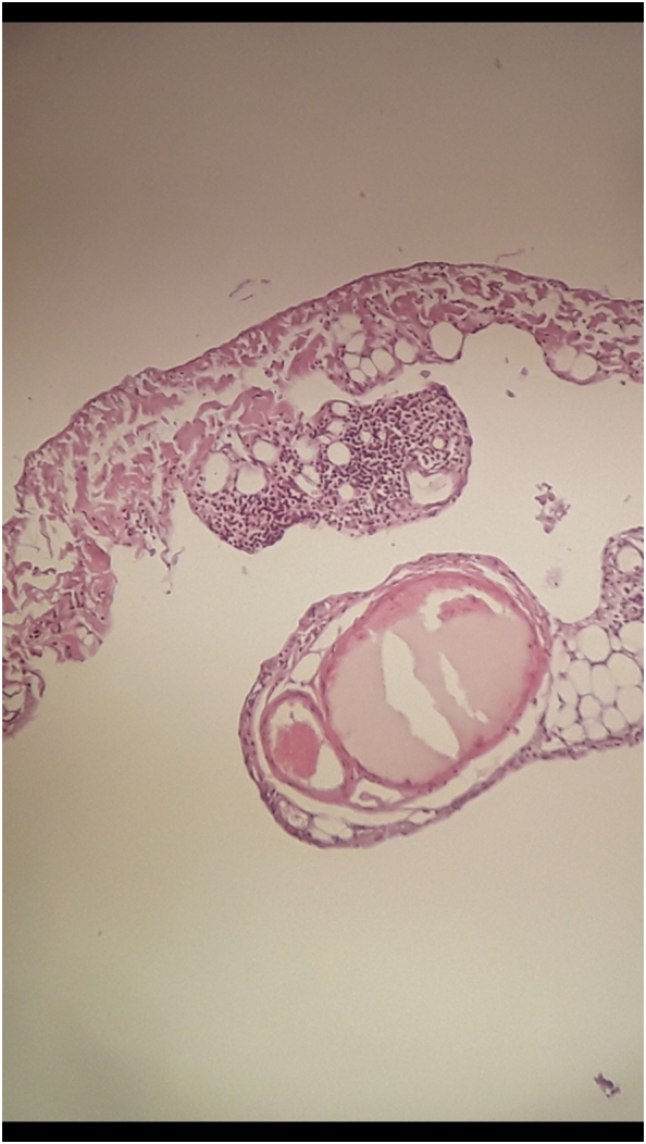
Case presentation: We present the case of a child who presented with a painless and non-obstructing abdominal mass. The mass was diagnosed and underwent complete surgical resection. Subsequent tissue analysis confirmed that the cyst was a lymphangioma.
Clinical discussion: These tumors should be considered in the differential diagnosis of cystic lesions in the abdomen, and the importance of performing complete surgical resection is emphasized.
Conclusion: The importance of complete surgical resection for mesenteric lymphangioma must be emphasized. Partial resection or aspiration should not be performed due to the risk of complications associated with these procedures and the increased risk of recurrence.
Keywords: Case report; Lymphangiomas; Lymphatic malformation; Pediatric surgery.
Copyright © 2024 The Authors. Published by Elsevier Ltd.. All rights reserved.
Conflict of interest statement
Declaration of competing interest The authors declare that they have no competing interests.
Figures


Similar articles
-
A rare pediatric case: Mesenteric cystic hygroma in a 5-year-old child.Int J Surg Case Rep. 2024 Nov;124:110435. doi: 10.1016/j.ijscr.2024.110435. Epub 2024 Oct 12. Int J Surg Case Rep. 2024. PMID: 39405765 Free PMC article.
-
A rare case of huge intra-abdominal cystic lymphangioma arising from rectovesical pouch; a case report.Int J Surg Case Rep. 2023 May;106:108275. doi: 10.1016/j.ijscr.2023.108275. Epub 2023 Apr 29. Int J Surg Case Rep. 2023. PMID: 37130478 Free PMC article.
-
Jejunal mesenteric lymphangioma treated by pancreaticoduodenectomy: A case report.Int J Surg Case Rep. 2020;77:165-169. doi: 10.1016/j.ijscr.2020.10.088. Epub 2020 Oct 22. Int J Surg Case Rep. 2020. PMID: 33161289 Free PMC article.
-
An acute presentation of pediatric mesenteric lymphangioma: a case report and literature overview.Acta Chir Belg. 2018 Oct;118(5):331-335. doi: 10.1080/00015458.2017.1379802. Epub 2017 Sep 19. Acta Chir Belg. 2018. PMID: 28927352 Review.
-
A rare case of giant mesenteric cystic lymphangioma of the small bowel in an adult: A case presentation and literature review.Acta Gastroenterol Belg. 2016 Sep-Dec;79(3):491-493. Acta Gastroenterol Belg. 2016. PMID: 28209109 Review.
Cited by
-
Giant mesenteric cysts: Clinical insights and diagnostic challenges.Int J Surg Case Rep. 2025 Feb;127:110941. doi: 10.1016/j.ijscr.2025.110941. Epub 2025 Jan 25. Int J Surg Case Rep. 2025. PMID: 39864218 Free PMC article.
-
Unusual retroperitoneal presentation of a Giant mesenteric cyst in a pediatric patient: Surgical case report.Int J Surg Case Rep. 2025 Sep;134:111775. doi: 10.1016/j.ijscr.2025.111775. Epub 2025 Aug 11. Int J Surg Case Rep. 2025. PMID: 40812034 Free PMC article.
References
-
- Manikoth P., Mangalore G.P., Megha V. Axillary cystic hygroma. J. Postgrad. Med. 2004;50(3):215–216. - PubMed
Publication types
LinkOut - more resources
Full Text Sources
