

Does CISS MRI Reliably Depict the Endolymphatic Duct in Children with and without Vestibular Aqueduct Enlargement?
- PMID: 38423746
- PMCID: PMC11288572
- DOI: 10.3174/ajnr.A8158
Does CISS MRI Reliably Depict the Endolymphatic Duct in Children with and without Vestibular Aqueduct Enlargement?
Abstract
Background and purpose: High-resolution CT is the mainstay for diagnosing an enlarged vestibular aqueduct (EVA), but MR imaging may be an appealing alternative, given its lack of ionizing radiation exposure. The purpose of this study was to determine how reliably MR imaging demonstrates the endolymphatic duct and endolymphatic duct enlargement in hearing-impaired children.
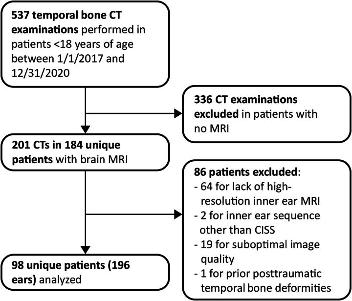
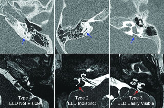
Materials and methods: We performed a retrospective review of temporal bone high-resolution CT and MR imaging of hearing-impaired children evaluated between 2017 and 2020. Vestibular aqueduct diameter was measured on high-resolution CT. The vestibular aqueducts were categorized as being enlarged (EVA+) or nonenlarged (EVA-) using the Cincinnati criteria. The endolymphatic ducts were assessed on axial high-resolution CISS MR imaging. We categorized endolymphatic duct visibility into the following: type 1 (not visible), type 2 (faintly visible), and type 3 (easily visible). Mixed-effect logistic regression was used to identify associations between endolymphatic duct visibility and EVA. Interreader agreement for the endolymphatic duct among 3 independent readers was assessed using the Fleiss κ statistic.
Results: In 196 ears from 98 children, endolymphatic duct visibility on MR imaging was type 1 in 74.0%, type 2 in 14.8%, and type 3 in 11.2%; 20.4% of ears were EVA+ on high-resolution CT. There was a significant association between EVA+ status and endolymphatic duct visibility (P < .01). Endolymphatic duct visibility was type 1 in 87.1%, type 2 in 12.8%, and type 3 in 0% of EVA- ears and type 1 in 22.5%, type 2 in 22.5%, and type 3 in 55.0% of EVA+ ears. The predicted probability of a type 3 endolymphatic duct being EVA+ was 0.997. There was almost perfect agreement among the 3 readers for distinguishing type 3 from type 1 or 2 endolymphatic ducts.
Conclusions: CISS MR imaging substantially underdiagnoses EVA; however, when a type 3 endolymphatic duct is evident, there is a >99% likelihood of an EVA.
© 2024 by American Journal of Neuroradiology.
Figures
Similar articles
-
MRI Evaluation of the Normal and Abnormal Endolymphatic Duct in the Pediatric Population: A Comparison with High-Resolution CT.AJNR Am J Neuroradiol. 2021 Oct;42(10):1865-1869. doi: 10.3174/ajnr.A7224. Epub 2021 Aug 26. AJNR Am J Neuroradiol. 2021. PMID: 34446455 Free PMC article.
-
Can magnetic resonance imaging provide clues to the inner ear functional status of enlarged vestibular aqueduct subjects with PDS mutation?Otol Neurotol. 2008 Aug;29(5):593-600. doi: 10.1097/MAO.0b013e318173033f. Otol Neurotol. 2008. PMID: 18665027
-
[Analysis of the correlation between radiological and audiological features of patients with enlarged vestibular aqueduct].Zhonghua Er Bi Yan Hou Tou Jing Wai Ke Za Zhi. 2019 Oct 7;54(10):734-740. doi: 10.3760/cma.j.issn.1673-0860.2019.10.006. Zhonghua Er Bi Yan Hou Tou Jing Wai Ke Za Zhi. 2019. PMID: 31606985 Chinese.
-
High jugular bulb with a diverticulum and vestibular aqueduct dehiscence: an anatomical variant to be aware in patients with hearing loss.Surg Radiol Anat. 2022 Jul;44(7):1041-1044. doi: 10.1007/s00276-022-02983-y. Epub 2022 Jul 16. Surg Radiol Anat. 2022. PMID: 35842486 Review.
-
[Enlarged vestibular aqueduct syndrome-dehiscence syndromes-honeycomb mastoid : Pathophysiology and evidence for clinical differentiation].HNO. 2020 May;68(5):336-343. doi: 10.1007/s00106-020-00837-w. HNO. 2020. PMID: 32347381 Review. German.
Cited by
-
Comparison of vestibular aqueduct visualization on computed tomography and magnetic resonance imaging in patients with Ménière's disease.BMC Med Imaging. 2024 Apr 22;24(1):93. doi: 10.1186/s12880-024-01275-8. BMC Med Imaging. 2024. PMID: 38649991 Free PMC article.
References
-
- Naganawa S, Koshikawa T, Iwayama E, et al. . MR imaging of the enlarged endolymphatic duct and sac syndrome by use of a 3D fast asymmetric spin-echo sequence: volume and signal-intensity measurement of the endolymphatic duct and sac and area measurement of the cochlear modiolus. AJNR Am J Neuroradiol 2000;21:1664–69 - PMC - PubMed
MeSH terms
LinkOut - more resources
Full Text Sources