. 2024 Feb 29;11(1):e001762.
doi: 10.1136/bmjresp-2023-001762.

Protective role of the HSP90 inhibitor, STA-9090, in lungs of SARS-CoV-2-infected Syrian golden hamsters

Affiliations

Protective role of the HSP90 inhibitor, STA-9090, in lungs of SARS-CoV-2-infected Syrian golden hamsters

Luiz Gustavo Teixeira Alves et al. BMJ Open Respir Res. .

Abstract

Introduction: The emergence of new SARS-CoV-2 variants, capable of escaping the humoral immunity acquired by the available vaccines, together with waning immunity and vaccine hesitancy, challenges the efficacy of the vaccination strategy in fighting COVID-19. Improved therapeutic strategies are urgently needed to better intervene particularly in severe cases of the disease. They should aim at controlling the hyperinflammatory state generated on infection, reducing lung tissue pathology and inhibiting viral replication. Previous research has pointed to a possible role for the chaperone HSP90 in SARS-CoV-2 replication and COVID-19 pathogenesis. Pharmacological intervention through HSP90 inhibitors was shown to be beneficial in the treatment of inflammatory diseases, infections and reducing replication of diverse viruses.

Methods: In this study, we investigated the effects of the potent HSP90 inhibitor Ganetespib (STA-9090) in vitro on alveolar epithelial cells and alveolar macrophages to characterise its effects on cell activation and viral replication. Additionally, the Syrian hamster animal model was used to evaluate its efficacy in controlling systemic inflammation and viral burden after infection.

Results: In vitro, STA-9090 reduced viral replication on alveolar epithelial cells in a dose-dependent manner and lowered significantly the expression of proinflammatory genes, in both alveolar epithelial cells and alveolar macrophages. In vivo, although no reduction in viral load was observed, administration of STA-9090 led to an overall improvement of the clinical condition of infected animals, with reduced oedema formation and lung tissue pathology.

Conclusion: Altogether, we show that HSP90 inhibition could serve as a potential treatment option for moderate and severe cases of COVID-19.

Keywords: COVID-19; inflammation; pneumonia; respiratory infection.


Conflict of interest statement

Competing interests: None declared.

Figures

Figure 1
Figure 1
Antiviral and anti-inflammatory properties of STA-9090 in vitro in infected AECs and alveolar macrophages. AECs, isolated from human lung tissue, were infected with MOI 0.5 of SARS-CoV-2 and treated with different concentrations of the HSP90 inhibitor STA-9090. STA-9090 concentrations ranged from 12.5 nM to a maximum of 800 nM on a log2 scale. Non-treated or non-infected cells serve as control. (A) Viral titers in supernatants of AECs infected with SARS-CoV-2 and treated with the indicated concentration of STA-9090. (B) Expression analysis of selected induced genes in AECs. Shown are z-scores of DESeq2 normalised counts comparing infected samples to uninfected control samples mock-treated or treated with different concentrations of STA-9090, as shown. Size of the circle and colours show differentiated expression. Alveolar macrophages were differentiated from lung tissue and infected with SARS-CoV-2 (MOI 0.5) prior to treatment with STA-9090. Mock infected cells, treated or non-treated, served as control. (C) Heatmaps of differently expressed genes in alveolar macrophages after infection with SARS-CoV-2 and treatment with 100 nM STA-9090. Uninfected cells, receiving STA-9090 or not, were used as controls. Genes related to IFN-mediated signalling pathway, type I IFN signalling, cellular-response to IFNy and cytokines were selected, as previously described. Columns represent samples and rows genes. Shown are z-scores of DESeq2-normalised data from two donors, analysed independently and colour scale ranges from blue (10% lower quantile) to red (10% upper quantile) of the selected genes. N=3 in A, C and n=2 in B. Unpaired t-test used for comparison in A. *p<0.05, **p<0.01. AECs, alveolar epithelial cells.
Figure 2
Figure 2
Treatment with STA-9090 does not affect viral loads in Syrian hamsters. (A) Syrian hamsters challenged with SARS-CoV-2 (1×10⁵ PFU), were treated with 25 mg/kg STA-9090 (GB) once, together with infection (group 1), or twice, at day 0 and at day 4 p.i. (group 2). Control group received same volume of dilutor at day 0 p.i. Analysis was made at day 3 (only for the control group and group 1), day 5 and day 7 p.i. (B) Body weight in percentage from the original weight was measured daily after infection of the animals with SARS-CoV-2. (C) Viral load was quantified by RT-qPCR analysis of viral genomic RNA (gRNA) copies detected in homogenised lung tissue and oropharyngeal swabs at days 3, 5 and 7 p.i. (D) Pharmacokinetics of STA-9090 quantified by mass spectrometry in the lung tissue or serum of Syrian hamsters infected with SARS-CoV-2. Shown is the number of days after STA-9090 application (first or second doses) in lung (left) and serum (right). Results are displayed as mean±SE and n=3. Comparisons were made with a one-way ANOVA test. ANOVA, analysis of variance. LOD, limit of detection.
Figure 3
Figure 3
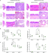
Lung tissue pathology scores are reduced in infected hamsters after treatment with STA-9090. Longitudinal sections of left lungs from Syrian hamsters after 5 days or 7 days infection with SARS-CoV-2 and receiving treatment of STA-9090. Treatment was performed at day 0 p.i. or at day 0 and again at day 4 p.i. and lung tissue was stained with H&E for histological comparisons (A). Shown are whole lung sections and magnification of the epithelial or endothelial layers. Lung pneumonia with oedema formation is seen in darker colours, due to the accumulation of fluids and cells. Representative histology is shown. Semiquantitative analysis of histological lesions quantified at days 3, 5 and 7 p.i. were scored and shown for comparisons. Lung affected area (B), alveolar oedema and AT2 cell hyperplasia (C) and lung endotheliitis (D) are shown. Results are displayed as mean±SE and n=3. Comparisons were made by a one-way ANOVA test. *p<0.05. ANOVA, analysis of variance.
Figure 4
Figure 4
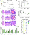
Early treatment with STA-9090 reduces inflammation and lung tissue pathology. (A) Syrian hamsters were challenged with SARS-CoV-2 (1×10⁵ PFU) and treated with 25 mg/kg STA-9090 (GB) at 48 h p.i. (GB 48 hours) or at 72 h p.i. (GB 72 hours). Control group received same volume of dilutor and analysis was made at day 5 p.i. (B) RT-qPCR analysis of viral genomic RNA (gRNA) copies detected in homogenised lung tissue was performed at day 5 p.i. (C) Longitudinal sections of left lungs of Syrian hamsters 5 days after infection with SARS-CoV-2 were stained with H&E for histological comparisons. Shown are whole lung sections, and sections focusing on epithelial cell layer and interstitial space. Lung pneumonia with oedema formation is seen in darker colours. Representative histology is shown. (D) Semiquantitative analysis of histological lesions quantified at day 5 p.i. were scored and shown for comparisons for lung affected area and alveolar oedema. Shown is the comparison between the groups receiving STA-9090 at day 2 or 3 p.i. and the control group. (E) Expression analysis of selected genes affected by infection and treatment with STA-9090 in blood samples of Syrian hamsters. Normalised expression values of selected cytokines are shown in logarithmic scale (left) and expression of genes related to tissue regeneration and repair are shown with z-score values of DESeq2 normalised counts (right). Size of the circle and colours show differentiated expression. Average values±SE are shown and n=4. Comparisons were made with a two-tailed Mann-Whitney test (B) and a one-way ANOVA test (D, E). *p<0.05. ANOVA, analysis of variance.

References

    1. WHO . Coronavirus disease (COVID-19) pandemic, . 2021.. Available: https://www.who.int/emergencies/diseases/novel-coronavirus-2019
    1. Wu F, Zhao S, Yu B, et al. . A new Coronavirus associated with human respiratory disease in China. Nature 2020;579:265–9. 10.1038/s41586-020-2008-3 - DOI - PMC - PubMed
    1. Wu Z, McGoogan JM. Characteristics of and important lessons from the Coronavirus disease 2019 (COVID-19) outbreak in China. JAMA 2020;323:1239. 10.1001/jama.2020.2648 - DOI - PubMed
    1. Osuchowski MF, Winkler MS, Skirecki T, et al. . The COVID-19 puzzle: Deciphering pathophysiology and phenotypes of a new disease entity. Lancet Respir Med 2021;9:622–42. 10.1016/S2213-2600(21)00218-6 - DOI - PMC - PubMed
    1. Liao M, Liu Y, Yuan J, et al. . Single-cell landscape of Bronchoalveolar immune cells in patients with COVID-19. Nat Med 2020;26:842–4. 10.1038/s41591-020-0901-9 - DOI - PubMed

Publication types

Supplementary concepts