. 2024 Feb 29;15(1):1884.
doi: 10.1038/s41467-024-45422-9.

High-throughput barcoding of nanoparticles identifies cationic, degradable lipid-like materials for mRNA delivery to the lungs in female preclinical models

Affiliations

High-throughput barcoding of nanoparticles identifies cationic, degradable lipid-like materials for mRNA delivery to the lungs in female preclinical models

Lulu Xue et al. Nat Commun. .

Abstract

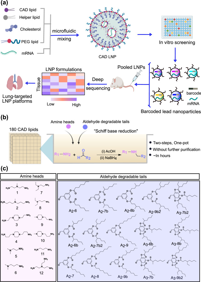
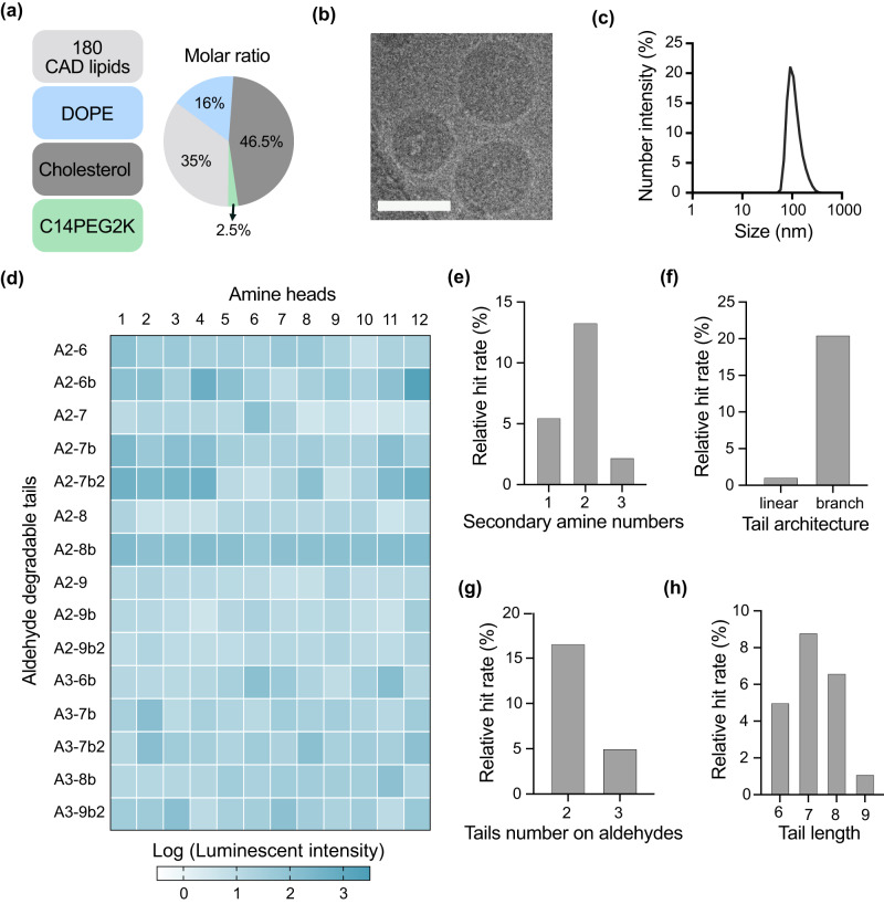
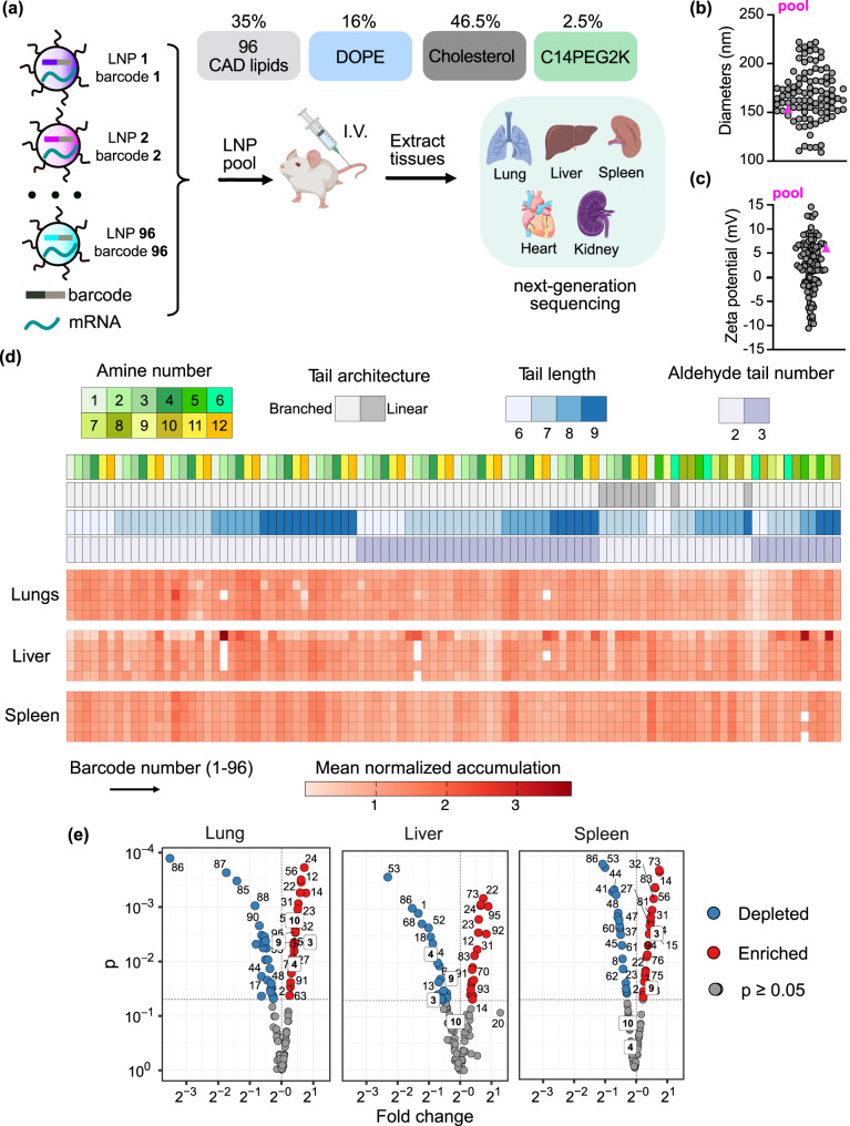
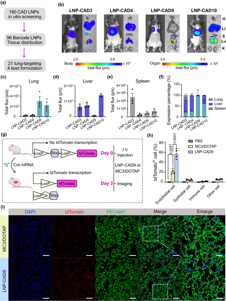
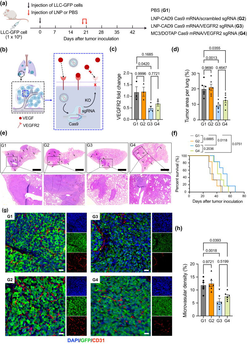
Lipid nanoparticles for delivering mRNA therapeutics hold immense promise for the treatment of a wide range of lung-associated diseases. However, the lack of effective methodologies capable of identifying the pulmonary delivery profile of chemically distinct lipid libraries poses a significant obstacle to the advancement of mRNA therapeutics. Here we report the implementation of a barcoded high-throughput screening system as a means to identify the lung-targeting efficacy of cationic, degradable lipid-like materials. We combinatorially synthesize 180 cationic, degradable lipids which are initially screened in vitro. We then use barcoding technology to quantify how the selected 96 distinct lipid nanoparticles deliver DNA barcodes in vivo. The top-performing nanoparticle formulation delivering Cas9-based genetic editors exhibits therapeutic potential for antiangiogenic cancer therapy within a lung tumor model in female mice. These data demonstrate that employing high-throughput barcoding technology as a screening tool for identifying nanoparticles with lung tropism holds potential for the development of next-generation extrahepatic delivery platforms.


Conflict of interest statement

L.X. and M.J.M. have filed a patent application on this research. D.W. is named on patents that describe the use of nucleoside-modified mRNA as a platform to deliver therapeutic proteins and vaccines. D.W. and M.G.A. are named on patents describing the use of lipid nanoparticles for nucleic acid delivery. The other authors declare no competing interests.

Figures

Fig. 1
Fig. 1. High-throughput LNP screening facilitates the discovery of cationic degradable (CAD) lipid-like materials for mRNA delivery to the lungs.
a CAD LNPs were initially formulated using a microfluidic mixing device by mixing nucleic acid with CAD lipids, helper lipid, cholesterol, and PEG-lipid. Following in vitro high-throughput screening, a series of CAD LNPs were selected and formulated to co-encapsulate b-DNA and mRNA, pooled, and systemically administered into mice, allowing for quantification of accumulation in each organ (heart, liver, spleen, lungs, and kidneys) using deep sequencing to identify CAD lipid candidates for lung-targeted mRNA delivery. b A combinatorial library of CAD lipids was chemically synthesized through “Schiff base reduction” by reacting amine heads and aldehyde degradable tails. c Overview of 12 amines cores and 15 aldehyde degradable tails used to synthesize 180 CAD lipids. a Created with BioRender.com.
Fig. 2
Fig. 2. Investigation of structure-activity relationship of CAD LNPs for firefly luciferase (FLuc) mRNA delivery in vitro.
a CAD LNP formulation parameters. CAD LNPs were formulated with one of 180 distinct CAD lipids, DOPE, cholesterol, and C14PEG2K at a molar ratio of 35:16:46.5:2.5, for a total of 180 distinct LNP formulations. b Representative cryogenic transmission electron microscopy (cryo-TEM) image of 3-A2−7b LNP morphology (n = 3 replicates). Scale bar: 100 nm. c Hydrodynamic size distribution of 3-A2-7b LNP obtained by dynamic light scattering (DLS). d A heatmap of luciferase expression following treatment of HeLa cells with CAD LNPs (10 ng luciferase mRNA, n ≥ 3 replicates). Relative luminescence unit (RLU) values of > 100 were classified as hits for hit rate calculation. e Relative hit rate of CAD LNPs with different secondary amine numbers. f Relative hit rate of CAD LNPs with different tail architectures. g Relative hit rate of CAD LNPs with different tail substitution numbers on each aldehyde. h Relative hit rate of CAD LNPs with different tail lengths. Source data are provided in the Source Data file.
Fig. 3
Fig. 3. In vivo structure-activity relationship analysis of 96 chemically distinct CAD lipids and organ tropism.
a Schematic illustration of barcoding approach to probe biodistribution profile of designed CAD LNPs. LNPs were formulated by pipette mixing to encapsulate barcoded DNA (b-DNA) and FLuc mRNA. Lipid phases consisted of one of 96 CAD lipids, DOPE, cholesterol, and C14PEG2K at a molar ratio of 35:16:46.5:2.5. LNP formulations were pooled together and administered systemically to C57BL/6 J female mice (n = 5). Tissues were isolated 6 h post-administration, DNA was extracted, and accumulation of b-DNAs was quantified by deep sequencing. I.V.: intravenous. b Hydrodynamic diameter of all administered CAD LNPs. The diameter of the LNP pool control (pink triangle symbol) falls within the range of the CAD LNPs composing the pool. c Zeta potential of all administered CAD LNPs. The zeta potential of the pooled LNP (pink triangle symbol) falls within the range of the CAD LNPs composing the pool. d Heatmap visualizing accumulation of CAD LNPs in different organs as measured by deep sequencing. Dark clusters represent higher relative accumulation of b-DNA in a specific tissue sample. Structural details of CAD lipids used in each LNP formulation are described above the heatmap. e Volcano plots summarizing enrichment analysis of barcodes in the lungs, liver, and spleen. The normalized accumulation of LNP formulation was compared to the aggregate LNP pool (i.e., basemean) using a two-sided Wilcoxon rank-sum test. False discovery rate was controlled using the Benjamini-Hochberg method. The exact P values from each comparison are provided in Supplementary Data 1. a Created with BioRender.com. Source data are provided in the Source Data file.
Fig. 4
Fig. 4. Validation of lead LNP formulations for mRNA delivery to the lungs of female mice.
a Four lead LNP formulations were discovered from a 180-CAD lipid library after high-throughput in vitro and in vivo screening. b Whole body and ex vivo imaging of luciferase expression mediated by LNP-CAD3, 4, 9, and 10 at 6 h post-injection (0.1 mg kg−1 FLuc mRNA, n = 3 mice). H: heart, Li: liver, S: spleen, Lu: lung, K: kidney. Quantification of luciferase expression in the lungs (c), liver (d), and spleen (e) using region-of-interest (ROI) analysis. f Relative luciferase expression in each measured organ. g Ai14 mice were treated with LNP-CAD9 or MC3/DOTAP LNP encapsulating Cre mRNA 3 days prior to analysis (0.3 mg kg−1, n = 4 mice). Lungs were digested and stained for quantifying cell populations for tdTomato expression. PBS was injected as negative control. I.V.: intravenous. h Proportion of tdTomato+ cells in the lung assessed by flow cytometry. i Representative immunostaining demonstrating signal overlap between tdTomato+ cells and the endothelial cell marker platelet endothelial cell adhesion molecule 1 (PECAM1). DAPI was used for nuclear staining. Scale bars: 100 µm for the lung section images and 30 µm for enlarged images. g Created with BioRender.com. Statistical significance in (h) was calculated using one-way analysis of variance (ANOVA), followed by Dunnett’s multiple comparison test. **P < 0.01; ***P < 0.001. Data are presented as mean ± s.e.m. Source data are provided in the Source Data file.
Fig. 5
Fig. 5. Antiangiogenic therapy through knockout  of VEGFR2 by LNPs in orthotropic lung cancer model.
a Schematic illustration of lung tumor implantation through i.v. injection of Lewis lung carcinoma cell lines expressing GFP (LLC-GFP) and treatment protocol of C57BL/6J female mice. On day 20 after tumor cell inoculation, mice were randomly assigned to four groups: PBS treated control (G1), LNP-CAD9 encapsulating Cas9 mRNA/scrambled sgRNA treatment (G2), LNP-CAD9 encapsulating Cas9 mRNA/VEGFR2 sgRNA treatment (G3), and MC3/DOTAP LNP encapsulating Cas9 mRNA/VEGFR2 sgRNA treatment (G4). Mice were treated every other day for a total of 2 doses (2.0 mg kg−1 of RNA per injection). Seven days after the last administration, 6 mice in each group were euthanized and their lungs were isolated for antiangiogenic analysis. The remaining mice were used for survival evaluation. b Antiangiogenic mechanism through LNP-mediated VEGFR2 knockout. LNPs encapsulating Cas9 mRNA/VEGFR2 sgRNA demonstrated the ability to reduce the expression of VEGFR2 in lung endothelial cells, which inhibited the VEGF-VEGFR2 pathway. KO: knockout. c RT-qPCR measurement of VEGFR2 level in different treatment groups (n = 3 mice for G1 and G2 groups; n = 4 mice for G3 and G4 groups). d Quantification of tumor areas per lung of different treatment groups (n = 6 mice). e Representative H&E staining of lung tissue after sacrifice (n = 6 mice). Arrows indicate tumor areas in the lungs. Scale bar: 1 mm. f Percent survival of mice under different treatments (n = 6 mice). g Representative immunostaining of tumor areas in the lungs. Endothelial cells were stained by CD31 antibody. DAPI was used for nuclear staining. Scale bar: 100 µm. h Microvascular density (MVD) in the tumor area under different treatments (n = 6 mice). a, b Created with BioRender.com. Statistical significance in (c), (d), and (h) was calculated using one-way analysis of variance (ANOVA), followed by Dunnett’s multiple comparison test. Statistical significance in (f) was calculated using a log-rank test. ***p < 0.001; **p < 0.01; *p < 0.05; p > 0.05, not significant. Data are presented as mean ± s.e.m. Source data are provided in the Source Data file.

Similar articles

Cited by

References

    1. Chaudhary N, Weissman D, Whitehead KA. mRNA vaccines for infectious diseases: principles, delivery and clinical translation. Nat. Rev. Drug. Discov. 2021;20:817–838. doi: 10.1038/s41573-021-00283-5. - DOI - PMC - PubMed
    1. Pardi N, Hogan MJ, Porter FW, Weissman D. mRNA vaccines—a new era in vaccinology. Nat. Rev. Drug. Discov. 2018;17:261–279. doi: 10.1038/nrd.2017.243. - DOI - PMC - PubMed
    1. Sahin U, Karikó K, Türeci O. mRNA-based therapeutics–developing a new class of drugs. Nat. Rev. Drug. Discov. 2014;13:759–780. doi: 10.1038/nrd4278. - DOI - PubMed
    1. Mendes BB, et al. Nanodelivery of nucleic acids. Nat. Rev. Methods. Primers. 2022;2:24. doi: 10.1038/s43586-022-00104-y. - DOI - PMC - PubMed
    1. Pastor F, et al. An RNA toolbox for cancer immunotherapy. Nat. Rev. Drug. Discov. 2018;17:751–767. doi: 10.1038/nrd.2018.132. - DOI - PubMed