. 2024 Feb 29;14(1):34.
doi: 10.1038/s41408-024-01002-0.

Dual T-cell constant β chain (TRBC)1 and TRBC2 staining for the identification of T-cell neoplasms by flow cytometry

Affiliations

Dual T-cell constant β chain (TRBC)1 and TRBC2 staining for the identification of T-cell neoplasms by flow cytometry

Pedro Horna et al. Blood Cancer J. .

Abstract

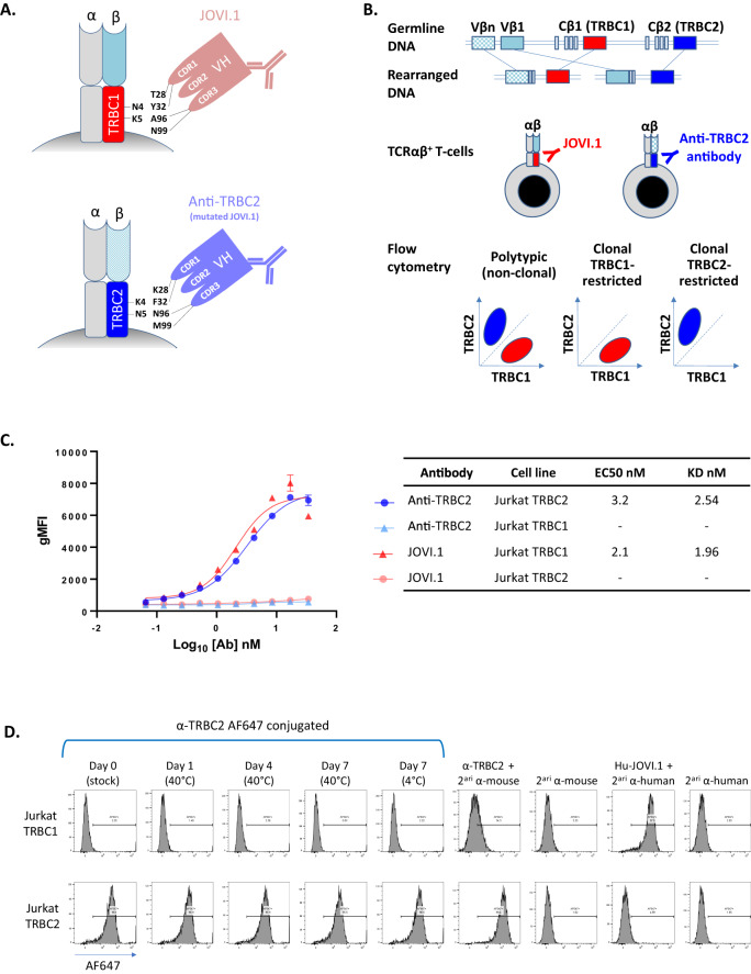
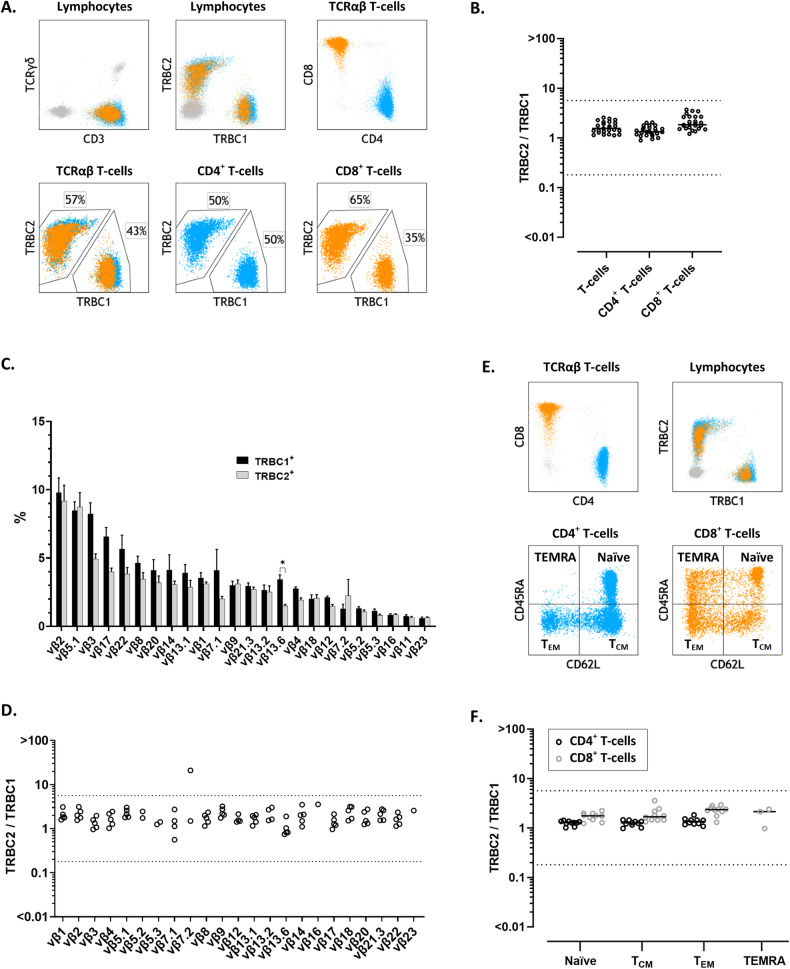
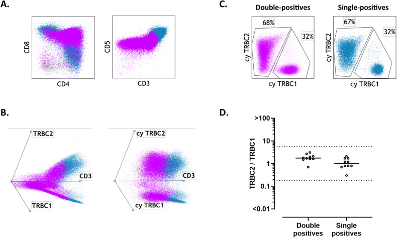
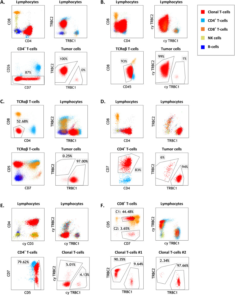
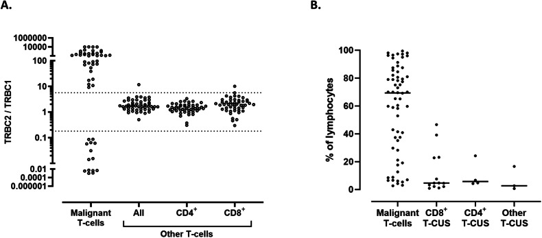
The diagnosis of leukemic T-cell malignancies is often challenging, due to overlapping features with reactive T-cells and limitations of currently available T-cell clonality assays. Recently developed therapeutic antibodies specific for the mutually exclusive T-cell receptor constant β chain (TRBC)1 and TRBC2 isoforms provide a unique opportunity to assess for TRBC-restriction as a surrogate of clonality in the flow cytometric analysis of T-cell neoplasms. To demonstrate the diagnostic utility of this approach, we studied 164 clinical specimens with (60) or without (104) T-cell neoplasia, in addition to 39 blood samples from healthy donors. Dual TRBC1 and TRBC2 expression was studied within a comprehensive T-cell panel, in a fashion similar to the routine evaluation of kappa and lambda immunoglobulin light chains for the detection of clonal B-cells. Polytypic TRBC expression was demonstrated on total, CD4+ and CD8+ T-cells from all healthy donors; and by intracellular staining on benign T-cell precursors. All neoplastic T-cells were TRBC-restricted, except for 8 cases (13%) lacking TRBC expression. T-cell clones of uncertain significance were identified in 17 samples without T-cell malignancy (13%) and accounted for smaller subsets than neoplastic clones (median: 4.7 vs. 69% of lymphocytes, p < 0.0001). Single staining for TRBC1 produced spurious TRBC1-dim subsets in 24 clinical specimens (15%), all of which resolved with dual TRBC1/2 staining. Assessment of TRBC restriction by flow cytometry provides a rapid diagnostic method to detect clonal T-cells, and to accurately determine the targetable TRBC isoform expressed by T-cell malignancies.


Conflict of interest statement

MP and PMM are inventors on a patent describing the use of TRBC1/2 for diagnosis and treatment of T-cell malignancies. MP and MF are inventors on a patent describing TRBC2 antibodies. Autolus Therapeutics owns patents claiming the use of TRBC1/2 for diagnosis. MP, MF, and ZA own stock in and are employees of Autolus Therapeutics. FTI is a former Autolus employee. The remaining authors declare no competing interests.

Figures

Fig. 1
Fig. 1. Strategic mutations on complementarity determining regions (CDR) of the JOVI.1 antibody result in switched specificity from T-cell receptor constant β chain (TRBC)1 to TRBC2, allowing for dual TRBC staining by flow cytometry.
A Simplified model of JOVI.1 antibody binding to TRBC1 (top), showing key amino acid residues on both molecules responsible for the discriminative binding to one isoform only. Rationally-designed antibody mutations on CDR1 (T28K and Y32F) and CDR3 (A96N and N99M) of JOVI.1’s variable heavy chain (VH) domain results in switched specificity to TRBC2 (bottom). B T-cell receptor αβ gene rearrangement showing the random selection of 1 of 2 mutually exclusive TRBC genes. Anti-TRBC1 (JOVI.1) and anti-TRBC2 antibodies can be utilized in conjunction to assess for TRBC-restriction by flow cytometry as a surrogate for T-cell clonality. C Half maximal effective concentration (EC50) measurements and dissociation constant (KD) estimations (non-linear regression one site binding analysis) of anti-TRBC2 (blue and light blue) or JOVI.1 (red and pink) binding to TRBC1-positive (triangles) or TRBC2-positive (circles) Jurkat cells; as assessed by geometric mean fluorescence intensity (gMFI) using an anti-mouse IgG fluorescent-labeled secondary antibody. D Flow cytometry histograms showing the specificity of the anti-TRBC2 antibody for TRBC2-positive (top) as compared to TRBC1-positive (bottom) Jurkat cells; and stability of an Alexa Fluor(AF)-647-conjugated anti-TRBC2 antibody. Secondary (2ari) antibodies were AF-647 conjugates. A humanized JOVI.1 antibody (Hu-JOVI.1) is also shown as control.
Fig. 2
Fig. 2. Dual staining for T-cell receptor constant β chain (TRBC)1 and TRBC2 demonstrates mutually exclusive and polytypic TRBC expression on benign T-cells and T-cell receptor variable β (TCR-Vβ) subsets.
A Representative peripheral blood flow cytometry findings on a healthy donor showing polytypic TRBC expression on total, CD4+ (cyan) and CD8+ (orange) T-cells. B TRBC2:TRBC1 ratios of total, CD4+ and CD8+ T-cells from 25 healthy donor’s peripheral blood specimens. Solid lines: medians. Dotted lines: thresholds for clonality. C TCR-Vβ repertoire by flow cytometry on gated TRBC1+ (black bars) and TRBC1- (gray bars) peripheral blood T-cells from five healthy donors, showing remarkably similar distributions. *p < 0.05. D TRBC2:TRBC1 ratios of each TCR-Vβ-positive T-cell subset from five healthy donors. Dotted lines depict thresholds for clonality. E Representative peripheral blood flow cytometry plots from a healthy donor showing distinct naïve, central memory (TCM), effector memory (TEM), and effector memory with reacquired CD45RA (TEMRA) subsets on the CD4+ (cyan) and CD8+ (orange) T-cell compartments. F TRBC2:TRBC1 ratios of gated naïve-memory CD4+ and CD8+ T-cell subsets from 10 healthy donors. Solid lines: medians. Dotted lines: thresholds for clonality.
Fig. 3
Fig. 3. Benign T-cell precursors show polytypic T-cell receptor constant β chain (TRBC) expression using dual cytoplasmic TRBC1 and TRBC2 staining.
A Representative immunophenotypic findings of a mediastinal mass involved by thymoma, showing benign CD4/CD8-double-positive T-cell precursors (violet) gradually acquiring surface CD3 expression as they mature into single-positive T-cells (blue). B Left: Double-positive T-cell precursors (violet) gradually acquire surface TRBC1 and TRBC2 expression while maturing into polytypic single-positive cells (blue). Right: Cytoplasmic TRBC1 and TRBC2 staining demonstrates polytypic TRBC expression on double-positive T-cell precursors. C Dual cytoplasmic TRBC1 and TRBC2 staining showing polytypic TRBC expression on double-positive and single-positive T-cell precursors. D TRBC2:TRBC1 ratios on double-positive and single-positive immature T-cells, using cytoplasmic dual staining on ten tissue biopsies with benign T-cell precursors.
Fig. 4
Fig. 4. Dual T-cell receptor constant β chain (TRBC)1 and TRBC2 staining by flow cytometry demonstrates TRBC restriction on gated malignant T-cells.
A Representative flow cytometry plots of peripheral blood involvement by Sezary syndrome (Sezary panel), showing a distinctly abnormal CD4+ T-cell subset (red) with loss of CD26 expression and TRBC2-restriction. Also shown are background polytypic CD4+ T-cells (cyan), CD8+ T-cells (orange), NK cells (gold) and B-cells (blue). B Peripheral blood involvement by T lymphoblastic leukemia/lymphoma (T-cell panel), showing an abnormal CD4-variable/CD8-dim T-cell population (red) that was surface CD3/TCR negative (data not shown) and on which TRBC2-restriction could be demonstrated by cytoplasmic (cy) TRBC1 and TRBC2 staining. C Inguinal lymph node involvement by cutaneous T-cell lymphoma, not otherwise specified, showing an expanded CD4/CD8-double-negative T-cell subset with TRBC1-restriction. D Cervical lymph node biopsy involved a CD4-positive peripheral T-cell lymphoma, showing a large subset of CD4+/CD7+ T-cells with TRBC1-restriction and absence of overt immunophenotypic aberrancies. E Peripheral blood from a patient with a lymphocytic variant of hypereosinophilic syndrome, showing an abnormal CD4+ T-cell subset negative for surface CD3 (not shown), positive for cytoplasmic CD3, and negative for surface and cytoplasmic TRBC. F Peripheral blood from a patient with Felty syndrome, showing 2 small CD8+ T-cell subsets with opposite TRBC-restriction, consistent with T-cell clones of uncertain significance.
Fig. 5
Fig. 5. Evaluation of T-cell receptor constant β chain (TRBC)2:TRBC1 ratio on T-cell subsets identifies malignant populations and small T-cell clones of uncertain significance (T-CUS).
A TRBC2:TRBC1 ratios of gated malignant T-cell populations from 52 clinical specimens with various T-cell neoplasms (excluding eight neoplasms lacking intracellular TRBC expression). Also shown are TRBC2:TRBC1 ratios of background (non-malignant) T-cells, including a blood sample with a small CD8+ T-cell clone of uncertain significance (outlier) in the setting of CD4+ cutaneous T-cell lymphoma. Dotted lines: thresholds for clonality. B Clone size of 62 T-cell neoplasms detected on 60 samples (expressed as a percentage of lymphocytes), compared to 20 T-CUS detected on 17 benign specimens.
Fig. 6
Fig. 6. Dual T-cell receptor constant β chain (TRBC)1/TRBC2 assessment resolves common artifacts encountered with TRBC1-only staining.
Specimens involved by cutaneous T-cell lymphoma (AC and F.), T-cell large granular lymphocytic leukemia (D, E), and samples from patients with no evidence of T-cell malignancy (GL), were studied using TRBC1-only staining (left) or dual TRBC1/TRBC2 staining (right). Neoplastic cells (red) with spurious dim expression using TRBC1-only staining are clearly resolved as TRBC1-restricted (AC) or TRBC2-restricted (DF) neoplasms using dual TRBC1/TRBC2 staining. On benign samples, spurious dim-expressing subsets (maroon) detected with TRBC1-only staining on CD8+ T-cells (G, H), CD4+ T-cells (I, J), or both (K, L), are completely resolved with dual TRBC1/TRBC2 staining.

References

    1. Jevremovic D, Olteanu H. Flow cytometry applications in the diagnosis of T/NK-cell lymphoproliferative disorders. Cytom B Clin Cytom. 2019;96:99–115. doi: 10.1002/cyto.b.21768. - DOI - PubMed
    1. Kroft SH, Harrington AM. How I diagnose mature T-cell proliferations by flow cytometry. Am J Clin Pathol. 2022;158:456–71. doi: 10.1093/ajcp/aqac079. - DOI - PubMed
    1. Tembhare PR, Chatterjee G, Chaturvedi A, Dasgupta N, Khanka T, Verma S, et al. Critical role of flow cytometric immunophenotyping in the diagnosis, subtyping, and staging of T-cell/NK-cell non-Hodgkin’s lymphoma in real-world practice: a study of 232 cases from a tertiary cancer center in India. Front Oncol. 2022;12:779230. doi: 10.3389/fonc.2022.779230. - DOI - PMC - PubMed
    1. Horna P, Olteanu H, Kroft SH, Harrington AM. Flow cytometric analysis of surface light chain expression patterns in B-cell lymphomas using monoclonal and polyclonal antibodies. Am J Clin Pathol. 2011;136:954–9. doi: 10.1309/AJCP3C2QZZBPTMLB. - DOI - PubMed
    1. Langerak AW, Groenen PJ, Bruggemann M, Beldjord K, Bellan C, Bonello L, et al. EuroClonality/BIOMED-2 guidelines for interpretation and reporting of Ig/TCR clonality testing in suspected lymphoproliferations. Leukemia. 2012;26:2159–71. doi: 10.1038/leu.2012.246. - DOI - PMC - PubMed